
|
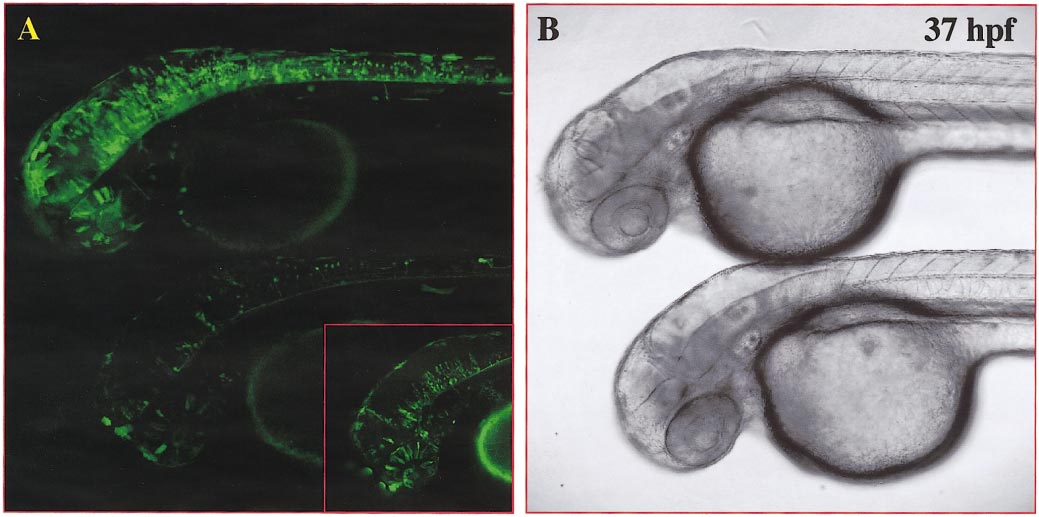
Fig. 3 Combined Gal4-VP16-activator/effector constructs result in strong EGFP expression in numerous cells of DNA-injected embryos. Single optical confocal microscopy section (lateral view) focusing on the nervous system of injected embryos at 37 hpf. Two embryos were embedded together in low-melting agarose to allow direct comparison of EGFP-expression levels and number of EGFP-expressing cells in a single optical section. Fluorescent (A pseudocolored in green) and transmitted light (B) images were taken at the same focal plane without moving the embryos. (A, B) EF-GVP-UG-injected embryos of the same batch. EGFP expression in EF-GVP-UG-injected embryos can vary strongly in intensity and frequency from a high number of embryonic GFP-expressing cells (A, upper embryo) to very few EGFP-expressing cells (A, lower embryo). Higher laser power and increased contrast, however, revealed a higher frequency of EGFP-expressing cells in the lower embryo than judged by the initial conditions (see A, inset).
Reprinted from Developmental Biology, 233(2), Köster, R.W. and Fraser, S.E., Tracing transgene expression in living zebrafish embryos, 329-346, Copyright (2001) with permission from Elsevier. Full text @ Dev. Biol.