
FIGURE
Fig. 6
- ID
- ZDB-FIG-080514-6
- Publication
- Kondo et al., 2001 - Dispersion of cyclin B mRNA aggregation is coupled with translational activation of the mRNA during zebrafish oocyte maturation
- Other Figures
- All Figure Page
- Back to All Figure Page
Fig. 6
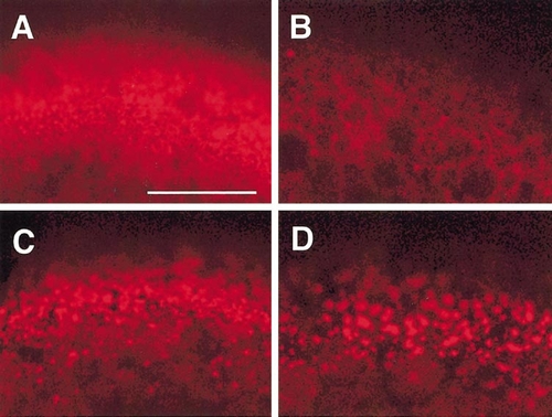
Microfilaments in full-grown immature (A), 17α,20β-DP-induced mature (B), and cytochalasin B-treated (C, D) oocytes, as revealed by confocal microscopy with rhodamine–phalloidin. Immature oocytes were treated with 1 (C) or 10 μg/ml (D) cytochalasin B. Dense meshwork of microfilaments is present in the cortical cytoplasm of immature oocytes (A) but not in mature oocytes (B). Cytochalasin B disrupts the microfilament meshwork partially at 1 μg/ml (C) and more extensively at 10 μg/ml (D). Scale, 50 μm. |
Expression Data
Expression Detail
Antibody Labeling
Phenotype Data
Phenotype Detail
Acknowledgments
This image is the copyrighted work of the attributed author or publisher, and
ZFIN has permission only to display this image to its users.
Additional permissions should be obtained from the applicable author or publisher of the image.
Reprinted from Developmental Biology, 229(2), Kondo, T., Kotani, T., and Yamashita, M., Dispersion of cyclin B mRNA aggregation is coupled with translational activation of the mRNA during zebrafish oocyte maturation, 421-431, Copyright (2001) with permission from Elsevier. Full text @ Dev. Biol.