
Fig. 4
- ID
- ZDB-FIG-080514-4
- Publication
- Kondo et al., 2001 - Dispersion of cyclin B mRNA aggregation is coupled with translational activation of the mRNA during zebrafish oocyte maturation
- Other Figures
- All Figure Page
- Back to All Figure Page
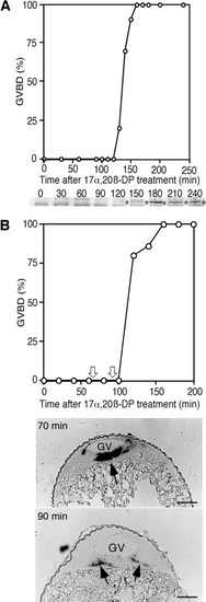
Cyclin B mRNA, cyclin B protein, and GVBD during 17α,20β-DP-induced oocyte maturation. (A) Time course of GVBD and cyclin B protein synthesis during oocyte maturation, indicating that both occur almost simultaneously 120 min after 17α,20β-DP treatment. Cyclin B proteins on the blot are indicated by asterisks. (B) Time course of GVBD and cyclin B mRNA dispersion. Oocytes collected at 70 and 90 min after 17α,20β-DP treatment (indicated by white arrows) were stratified by centrifugation in a density gradient of Ficoll. Aggregation is seen in an oocyte collected at 70 min, while it was diffused in the oocytes at 90 min just prior to the initiation of cyclin B synthesis and GVBD. Black arrows indicate cyclin B mRNA. GV, germinal vesicle. Scale, 100 μm. |
Reprinted from Developmental Biology, 229(2), Kondo, T., Kotani, T., and Yamashita, M., Dispersion of cyclin B mRNA aggregation is coupled with translational activation of the mRNA during zebrafish oocyte maturation, 421-431, Copyright (2001) with permission from Elsevier. Full text @ Dev. Biol.