
FIGURE
Fig. 3
- ID
- ZDB-FIG-080514-3
- Publication
- Kondo et al., 2001 - Dispersion of cyclin B mRNA aggregation is coupled with translational activation of the mRNA during zebrafish oocyte maturation
- Other Figures
- All Figure Page
- Back to All Figure Page
Fig. 3
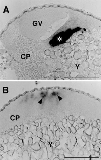
Cyclin B mRNA in immature (A) and mature (B) oocytes stratified by centrifugation on a Ficoll density gradient. The centrifugation segregated the oocyte cytoplasm (CP), including the germinal vesicle (GV), from the metaplasm consisting mainly of yolk (Y). Although cyclin B mRNA was seen in the cytoplasmic layer of both immature and mature oocytes, it exists as an aggregated form (asterisk) in immature oocytes and as indistinct matter (arrowheads) in mature oocytes. Scale, 100 μm. |
Expression Data
Expression Detail
Antibody Labeling
Phenotype Data
Phenotype Detail
Acknowledgments
This image is the copyrighted work of the attributed author or publisher, and
ZFIN has permission only to display this image to its users.
Additional permissions should be obtained from the applicable author or publisher of the image.
Reprinted from Developmental Biology, 229(2), Kondo, T., Kotani, T., and Yamashita, M., Dispersion of cyclin B mRNA aggregation is coupled with translational activation of the mRNA during zebrafish oocyte maturation, 421-431, Copyright (2001) with permission from Elsevier. Full text @ Dev. Biol.