
Fig. 7
- ID
- ZDB-FIG-080422-28
- Publication
- Kurita et al., 2003 - Suppression of lens growth by alphaA-crystallin promoter-driven expression of diphtheria toxin results in disruption of retinal cell organization in zebrafish
- Other Figures
- All Figure Page
- Back to All Figure Page
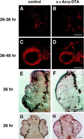
Cell cycle progression of zαAcry-DTA-expressing embryos examined by analyzing the incorporation of BrdU and anti Histone H3 antibody. BrdU was injected into the yolk of control (A, C) and zαAcry-DTA-expressing (B, D) embryos at 26 (A, B) or 36 (C, D) hpf, and the embryos were harvested at 36 (A, B) or 48 (C, D) hpf. Incorporated BrdU was visualized with Cy3-conjugated second antibody, and photos were taken under observation with an Axioplan microscope (Carl Zeiss). Scale bars, 50 μm. M-phase cells are visualized by the staining with anti-phospho Histone H3 antibody (E–H). Frozen sections of control (E, G) or zαAcry-DTA-expressing (F, H) embryos at 36 hpf were immunostained with anti-phospho Histone H3. Signals are visualized by using an ABC kit. (E, G) and (F, H) are different magnified pictures of different samples. Scale bars, 50 μm. |
Reprinted from Developmental Biology, 255(1), Kurita, R., Sagara, H., Aoki, Y., Link, B.A., Arai, K.-I., and Watanabe, S., Suppression of lens growth by alphaA-crystallin promoter-driven expression of diphtheria toxin results in disruption of retinal cell organization in zebrafish, 113-127, Copyright (2003) with permission from Elsevier. Full text @ Dev. Biol.