
Fig. 2
- ID
- ZDB-FIG-080422-23
- Publication
- Kurita et al., 2003 - Suppression of lens growth by alphaA-crystallin promoter-driven expression of diphtheria toxin results in disruption of retinal cell organization in zebrafish
- Other Figures
- All Figure Page
- Back to All Figure Page
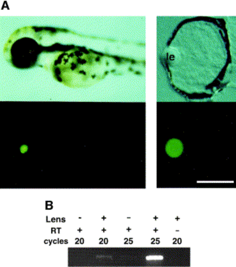
Zebrafish αA-crystallin promoter expressed EGFP lens-specific manner. (A) zαAcry-EGFP plasmid was injected into zebrafish eggs at the one- or two-cell stage, and lateral viewes under a dissection microscope are shown (left panel). An upper panel shows a view of a live embryo at 54 hpf, and a lower panel shows EGFP fluorescence image. Central transverse frozen sections were examined, and Normarsky image (right upper panel) and EGFP fluorescence (right lower panel) are shown. “le” indicates lens. Scale, 50 μM. (B) RT-PCR pattern of EGFP. EGFP-expressing embryos were collected and divided into two groups. For one group, the lenses were surgically removed; and for the other group, they were kept intact. Embryos were then decapitated, their heads were collected (more than 70 embryos for each group), and mRNA was prepared. RT-PCR of EGFP was done and visualized by agarose gel electrophoresis. (C) Alignment of the proximal 5′ region of zebrafish, chick, and mouse αA-crystallin gene. Conserved nucleotides were hatched, and cis-elements reported in chick (under the sequence) and mouse (above the sequence) are indicated (Cvekl and Piatigorsky 1996 and Ilagan et al 1999). (D) Summary of results of a series of deletion mutants from the 5′ side of zebrafish αA-crystallin. A series of deletion mutants of αA-crystallin promoter-EGFP was injected fertilized eggs at the one- or two-cell stage. The microinjected embryos were examined under a fluorescence dissection microscope. Square and circle indicate highly conserved regions. With chick and mouse promoters, it was reported that transcription factor binding sites were clustered in these region. |
Reprinted from Developmental Biology, 255(1), Kurita, R., Sagara, H., Aoki, Y., Link, B.A., Arai, K.-I., and Watanabe, S., Suppression of lens growth by alphaA-crystallin promoter-driven expression of diphtheria toxin results in disruption of retinal cell organization in zebrafish, 113-127, Copyright (2003) with permission from Elsevier. Full text @ Dev. Biol.