
Fig. 5
- ID
- ZDB-FIG-080422-26
- Publication
- Kurita et al., 2003 - Suppression of lens growth by alphaA-crystallin promoter-driven expression of diphtheria toxin results in disruption of retinal cell organization in zebrafish
- Other Figures
- All Figure Page
- Back to All Figure Page
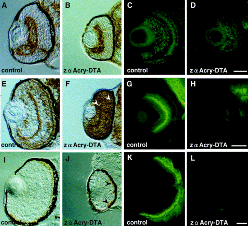
Immunohistochemical analysis of the sectioned zebrafish embryo eye with various retinal differentiation markers. (A, B) Retinal ganglion cells, stained with anti-neurolin (DM-GRASP) antibody (zn-5). (C, D) Retinal ganglion cells, inner nuclear layer, stained with anti-Islet 1. (E, F) Inner nuclear layer and outer plexiform layer, stained with Zns-2. (G, H) Amacrine and horizontal cells, stained with anti-syntaxin antibody. (I, J) Cone cell stained with Zpr-1. (K, L) Rod cells, stained with Zpr-3. Zebrafish eggs were injected with zαA-crystallin-DTA or control plasmid and central, transverse frozen sections were made at 48 (A, B, E, F), 54 (C, D, G, H), 72 (I, J), or 96 (K, L) h after fertilization. Immunohistochemistry was done, and staining was visualized by use of an ABC kit (A, B, E, F, I, J) or alexa 488 (C, D, G, H, K, L). Scale bars, 50 μm. |
Reprinted from Developmental Biology, 255(1), Kurita, R., Sagara, H., Aoki, Y., Link, B.A., Arai, K.-I., and Watanabe, S., Suppression of lens growth by alphaA-crystallin promoter-driven expression of diphtheria toxin results in disruption of retinal cell organization in zebrafish, 113-127, Copyright (2003) with permission from Elsevier. Full text @ Dev. Biol.