
FIGURE
Fig. 6
- ID
- ZDB-FIG-080422-27
- Publication
- Kurita et al., 2003 - Suppression of lens growth by alphaA-crystallin promoter-driven expression of diphtheria toxin results in disruption of retinal cell organization in zebrafish
- Other Figures
- All Figure Page
- Back to All Figure Page
Fig. 6
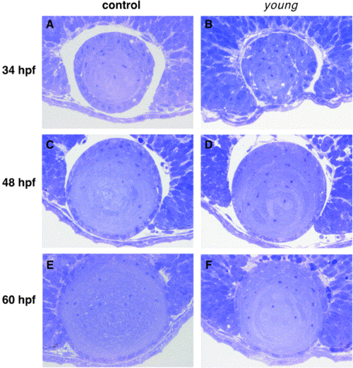
The lens development of young mutant. The morphology of lens of young mutant and controls at 34, 48, and 60 hpf were analyzed under light microscopy. Toluidine blue-stained sections through control (left panels) and young mutant (right panels) eyes at 34 (A, B), 48 (C, D), and 60 (E, F) hpf are shown. |
Expression Data
Expression Detail
Antibody Labeling
Phenotype Data
Phenotype Detail
Acknowledgments
This image is the copyrighted work of the attributed author or publisher, and
ZFIN has permission only to display this image to its users.
Additional permissions should be obtained from the applicable author or publisher of the image.
Reprinted from Developmental Biology, 255(1), Kurita, R., Sagara, H., Aoki, Y., Link, B.A., Arai, K.-I., and Watanabe, S., Suppression of lens growth by alphaA-crystallin promoter-driven expression of diphtheria toxin results in disruption of retinal cell organization in zebrafish, 113-127, Copyright (2003) with permission from Elsevier. Full text @ Dev. Biol.