
Fig. 5
- ID
- ZDB-FIG-080417-25
- Publication
- Kotani et al., 2008 - misty somites, a maternal effect gene identified by transposon-mediated insertional mutagenesis in zebrafish that is essential for the somite boundary maintenance
- Other Figures
- All Figure Page
- Back to All Figure Page
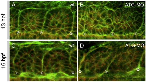
The mys-deficiency impaired epithelialization of somitic cells. (A–D) Confocal images of embryos stained with the anti-γ-Tubulin antibody (red) and Phalloidin (green). Dorsal views of a wild type embryo (A, C) and an ATG-MO injected embryo (B, D) at 13 hpf (the 6-somite stage) (A, B) or at 16 hpf (the 14-somite stage) (C, D). Asterisks indicate the positions of the somite boundaries. In wild type, somitic cells adjacent to the boundaries show a columnar shape and apical localization of the centrosomes. In the ATG-MO injected embryo, the somitic cells do not show the columnar shape and the centrosomes are distributed randomly in the cytoplasm. |
| Fish: | |
|---|---|
| Knockdown Reagent: | |
| Observed In: | |
| Stage Range: | 5-9 somites to 14-19 somites |
Reprinted from Developmental Biology, 316(2), Kotani, T., and Kawakami, K., misty somites, a maternal effect gene identified by transposon-mediated insertional mutagenesis in zebrafish that is essential for the somite boundary maintenance, 383-396, Copyright (2008) with permission from Elsevier. Full text @ Dev. Biol.