
Fig. 5
- ID
- ZDB-IMAGE-080417-32
- Publication
- Kotani et al., 2008 - misty somites, a maternal effect gene identified by transposon-mediated insertional mutagenesis in zebrafish that is essential for the somite boundary maintenance
- All Figures
- Figures for Kotani et al., 2008
|
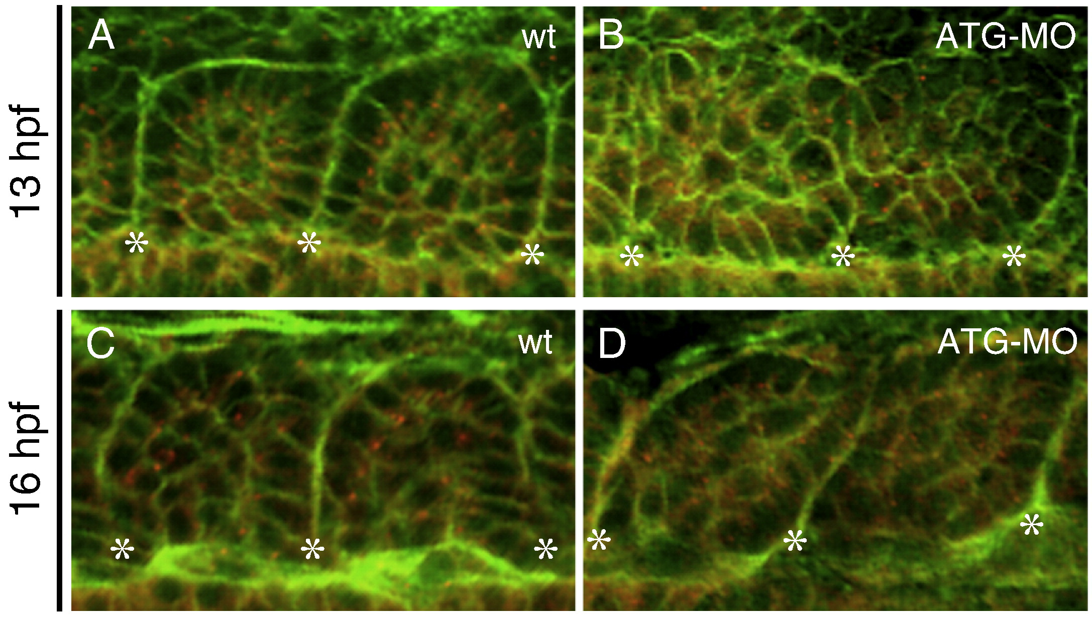
Fig. 5 The mys-deficiency impaired epithelialization of somitic cells. (A–D) Confocal images of embryos stained with the anti-γ-Tubulin antibody (red) and Phalloidin (green). Dorsal views of a wild type embryo (A, C) and an ATG-MO injected embryo (B, D) at 13 hpf (the 6-somite stage) (A, B) or at 16 hpf (the 14-somite stage) (C, D). Asterisks indicate the positions of the somite boundaries. In wild type, somitic cells adjacent to the boundaries show a columnar shape and apical localization of the centrosomes. In the ATG-MO injected embryo, the somitic cells do not show the columnar shape and the centrosomes are distributed randomly in the cytoplasm.
Reprinted from Developmental Biology, 316(2), Kotani, T., and Kawakami, K., misty somites, a maternal effect gene identified by transposon-mediated insertional mutagenesis in zebrafish that is essential for the somite boundary maintenance, 383-396, Copyright (2008) with permission from Elsevier. Full text @ Dev. Biol.