
FIGURE
Fig. 5
- ID
- ZDB-FIG-080402-28
- Publication
- De Mazière et al., 2008 - Egfl7 knockdown causes defects in the extension and junctional arrangements of endothelial cells during zebrafish vasculogenesis
- Other Figures
- All Figure Page
- Back to All Figure Page
Fig. 5
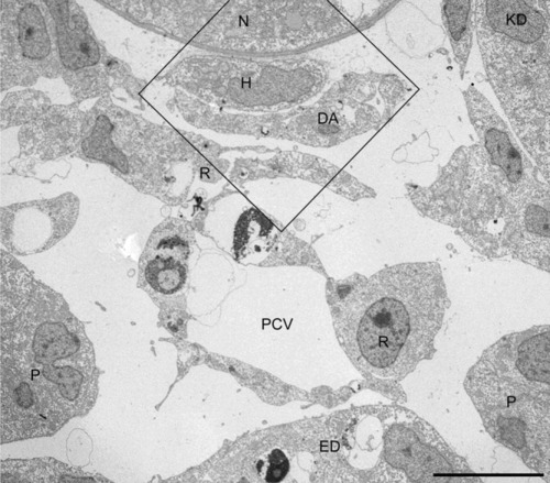
Altered positions of endothelial cells (ECs) in the axial vascular area of the trunk of Egfl7 KD embryos at 24 hours postfertilization (hpf). Cross-section of the vascular area. Dorsal aorta (DA), without lumen, and irregularly shaped posterior cardinal vein (PCV) are still associated with hypochord and endoderm, respectively. The box is enlarged in Figure 6. ED, endoderm; H, hypochord; N, notochord; P, pronephric duct; R, red blood cell precursor. Scale bar = 10 μm. |
Expression Data
Expression Detail
Antibody Labeling
Phenotype Data
| Fish: | |
|---|---|
| Knockdown Reagent: | |
| Observed In: | |
| Stage: | Prim-5 |
Phenotype Detail
Acknowledgments
This image is the copyrighted work of the attributed author or publisher, and
ZFIN has permission only to display this image to its users.
Additional permissions should be obtained from the applicable author or publisher of the image.
Full text @ Dev. Dyn.