
Fig. 3
- ID
- ZDB-FIG-080402-26
- Publication
- De Mazière et al., 2008 - Egfl7 knockdown causes defects in the extension and junctional arrangements of endothelial cells during zebrafish vasculogenesis
- Other Figures
- All Figure Page
- Back to All Figure Page
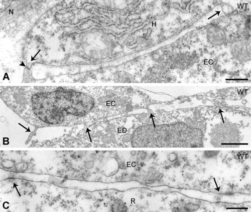
Heterologous cell interactions in the axial vascular area in the trunk of control embryos. A,B: Contact zone of dorsal aorta-endothelial cells (ECs) with the hypochord (A) and (B) of posterior cardinal vein-ECs with the endoderm. In both A and B, the cells contact each other by means of cellular extensions (arrows) approaching the opposed cell to a minimal distance. Arrowhead in A, gap junction. C: Between ECs and red blood cell precursors, deposits of extracellular material (arrows) are present at irregular distances. ED, endoderm; H, hypochord; N, notochord sheath; R, red blood cell precursor. Scale bars = 500 nm in A, 2 μm in B, 200 nm in C. |