
Fig. 1
- ID
- ZDB-FIG-080402-24
- Publication
- De Mazière et al., 2008 - Egfl7 knockdown causes defects in the extension and junctional arrangements of endothelial cells during zebrafish vasculogenesis
- Other Figures
- All Figure Page
- Back to All Figure Page
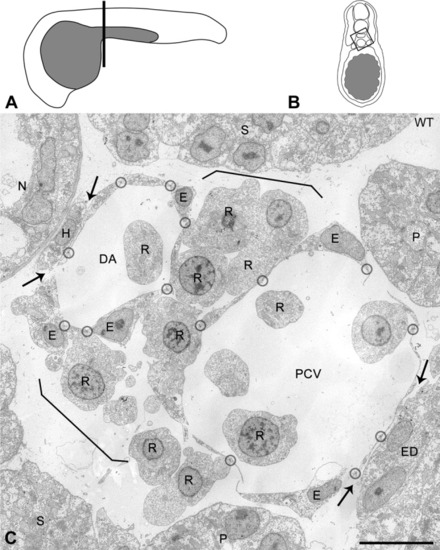
Cellular organization of the axial vascular area in the trunk of a 24 hours postfertilization (hpf) control embryo, viewed in cross-section. A: Scheme of a 24 hpf embryo with the position (line) of cross-sections. B: Scheme of a cross-section. Box: area analyzed by electron microscopy (EM). C: Axial vascular area of control (WT) embryo. Characteristically, the most dorsal endothelial cells (ECs) of the dorsal aorta (DA) are intimately associated with the hypochord (H), whereas the ventral part of the posterior cardinal vein (PCV) is associated with the endoderm (ED; arrows). Note that the extremely flattened ECs of the DA and PCV are connected by tiny contact zones (encircled), containing tight junctions and sometimes a desmosome or gap junction (see higher magnification in Fig. 2). Cells with morphological characteristics of red blood cell precursors (R) are observed in the vessel lumens and form a layer (between brackets) in between DA and PCV. E, nucleus of EC; N, notochord; P, pronephric duct; S, somite. Scale bar = 10 μm. |