
Fig. 2
- ID
- ZDB-FIG-080402-25
- Publication
- De Mazière et al., 2008 - Egfl7 knockdown causes defects in the extension and junctional arrangements of endothelial cells during zebrafish vasculogenesis
- Other Figures
- All Figure Page
- Back to All Figure Page
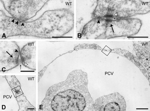
Endothelial cell-cell interactions in the axial vessels in the trunk of control embryos. A,B: Representative high magnification images of endothelial cell-endothelial cell (EC-EC) contact zones containing (A) only a tight junction consisting of a few plasma membrane fusion points (arrowhead), or (B) a tight junction (arrowhead) flanked by a desmosome (arrow). Note the tiny densities on the intercellular filamentous material at the central plane (small arrows) and the well-delineated dense plaques apposed to the plasma membranes (open arrowheads). C-E: EC-EC contact zone with tight junction (arrowhead) and desmosome (arrow), enlarged from boxed areas in D and E, showing the location of the desmosome between posterior cardinal vein (PCV) -ECs at a position luminal from the tight junction. Scale bars = 200 nm in A,B,D; 100 nm in C; 2 μm in E. |