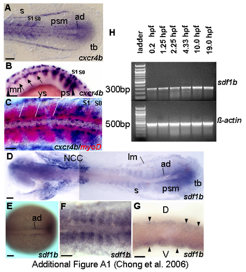
The dynamic expression of cxcr4b and sdf1b during segmentation. Dorsal views (A,C-F) and lateral views (B,G). (A) cxcr4b is expressed in the tailbud region, adaxial cells, paraxial mesoderm, 13.5 h. (B) Expression of cxcr4b is reduced as differentiation proceed, strong expression is in forming and newly formed somites, 18 h. (C) Two color in situ for myoD (red) and cxcr4b (blue) reveals partial overlapping expression of cxcr4b and myoD, 14 h. White lines demarcate the somite boundaries. (E) sdf1b transcription starts early in the adaxial cells, 10 h. (D,F) Expression in somites is relatively weak; some part of adaxial and paraxial mesoderm express sdf1b at 14 h and 14.5 h respectively. (G) sdf1b transcription localizes in dorsal and ventral regions of somites as indicated by black arrowheads, 16.5 h. (H) Reverse transcription (RT)-PCR detects continuous presence of transcript of sdf1b during early development. sdf1b transcript is present before mid-blastula transition (MBT). To confirm results, products were sequenced. β-actin was used as positive control. -RT control using β-actin primers without addition of reverse transcriptase, no band was detected (data not shown). Abbreviations: ad – adaxial cells; d – dorsal; lm – lateral mesoderm; mn – motoneurons; ncc – neural crest cells; ps – presomite; psm – presomitic mesoderm; s – somite; S0 – forming somite; S1 – newly formed somite; tb – tailbud; v – ventral; ys – yolk sac. Scale bars = 50 μm.
|
