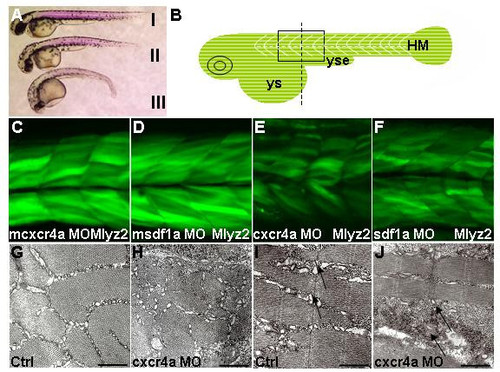
Formation of fast muscle requires Cxcr4a and Sdf1a. Lateral views (A-F), cross-section (G,H), sagittal section (I,J) and dorsal views. (A) Birefringence revealed by polarized light in cxcr4a (II) and sdf1a (III) morphants was reduced compared to control (I), 30 h. (B) Schematic illustrating black box region used for imaging. Start of yolk sac extension as a guide for the center of frame, indicated by dashed line in diagram of zebrafish embryo. (C-F) Single confocal images taken at level of the somite boundary as a guide of depth. Myosin light chain transgenic line, 51 h. (C,D) mcxcr4a (n = 87/87) and msdf1a (n = 31/35) morphants developed normally. (E,F) In representative cxcr4a (n = 63/71) and sdf1a (n = 73/82) morphants, reduction of GFP signal was observed. (G-J) Transmission electron micrograph of cross (G,H) and sagittal (I,J) sections in trunk region of control and cxcr4a morphants respectively, 36 h. A representative cxcr4a morphant clearly shows a reduction in muscle fibrils. (I,J) Black arrows indicate lack or absence of sarcoplasmic reticulum and muscle fibers in some areas of cxcr4a morphant. For clarity, this region of section (J) was selected where there are at least some muscle fibers. Abbreviations: HM – horizontal myoseptum; ys – yolk sac and yse – yolk sac extension. Scale bars = 500 nm.
|
