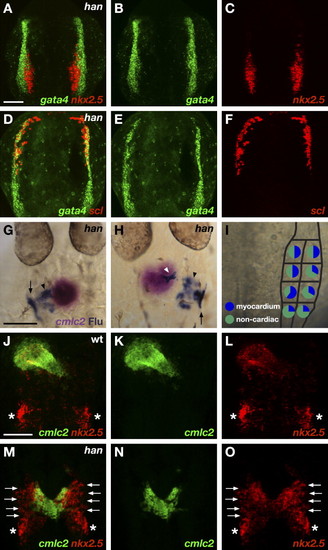
Hand2 Potentiates Myocardial Differentiation (A–F) ALPM territories appear to be established normally in han mutant embryos. Two-color fluorescent in situ hybridization of han mutants at the 7 somite stage, dorsal views, anterior to the top. The scale bar represents 100 μm. (A) Overlay of gata4 (B) and nkx2.5 (C) expression. (D) Overlay of gata4 (E) and scl (F) expression. Expression patterns of gata4, nkx2.5, and scl divide the han mutant ALPM into normally proportioned rostral, medial, and lateral regions. (G–I) Fate map of the caudal ALPM in han mutants indicates that Hand2 is required to facilitate effective differentiation of myocardial progenitors from medial and lateral ALPM territories. (G and H) Dorsal views of han mutant embryos at 30 hpf, anterior to the top. In situ hybridization indicates expression of cmlc2 (magenta) in cardiomyocytes, and immunohistochemistry for uncaged fluorescein (blue) indicates progeny of labeled cells. In han mutants, labeled cells from caudal ALPM can give rise to cardiomyocytes (white arrowhead; [H]) but also frequently become seemingly undifferentiated cells (black arrowheads; [G and H]) flanking the cardiomyocytes. Labeled cells also contribute to pharyngeal pouches (arrows; [G and H]). (I) Fate map of han mutant caudal ALPM. For each region tested, pie charts depict the proportions of experimental embryos producing labeled myocardial progeny (blue) and only noncardiac labeled progeny (green). Myocardial progenitors were found in all regions examined, but the frequency with which labeled han mutant ALPM produced cardiomyocytes was reduced, relative to wild-type, throughout both medial and lateral territories (compare to Figure 3B). See Table 1 for data reflected in schematic. (J–O) han mutants exhibit ineffective myocardial differentiation. Two-color fluorescent in situ hybridization at 24 hpf, dorsal views, anterior to the top. The scale bar represents 100 μm. (J and M) Overlays of cmlc2 (K and N) and nkx2.5 (L and O) expression. In wild-type embryos (J–L), cmlc2 and nkx2.5 are both expressed throughout the heart tube. Additional expression of nkx2.5 marks the forming pharyngeal pouches (asterisks). (M–O) In han mutants, a reduced number of cardiomyocytes span the midline. Undifferentiated, nkx2.5-expressing ALPM cells (arrows) flank the cardiomyocytes.
|
