
Fig. 3
- ID
- ZDB-FIG-070822-64
- Publication
- Schoenebeck et al., 2007 - Vessel and blood specification override cardiac potential in anterior mesoderm
- Other Figures
- All Figure Page
- Back to All Figure Page
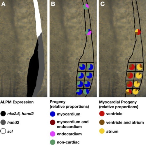
Fate Map of Cardiac Progenitors within ALPM Territories. Schematics depicting the progenitor composition of medial, lateral, and rostral ALPM territories. Dorsal views are of the right side of the ALPM, anterior to the top. (A) Schematic of gene expression patterns that distinguish medial (black), lateral (gray), and rostral (white) ALPM territories, as in Figure 1L. (B) Myocardial progenitors arise from medial and lateral territories and are only rarely detected in rostral territory. In contrast, endocardial progenitors arise from rostral territory. For each region tested, pie charts depict the proportions of experimental embryos exhibiting a particular type of labeled progeny. Colored pie pieces represent embryos that produced labeled myocardial progeny (blue), labeled endocardial progeny (pink), both myocardial and endocardial labeled progeny (maroon), and only noncardiac labeled progeny (green). Data shown are a merger of experiments from the left and right sides of the embryo; our data did not reveal any left-right asymmetry of the fate map. (C) Ventricular and atrial myocardial progenitors appear spatially organized within the caudal ALPM, with ventricular progenitors tending to originate medially and atrial progenitors tending to originate laterally. Pie charts depict the chamber contributions of labeled cells in the subset of experimental embryos containing labeled myocardial progeny. Colored pie pieces represent embryos that produced labeled ventricular cardiomyocytes (red), labeled atrial cardiomyocytes (yellow), and both ventricular and atrial cardiomyocytes (brown). See Table 1 for data reflected in schematics. |
Reprinted from Developmental Cell, 13(2), Schoenebeck, J.J., Keegan, B.R., and Yelon, D., Vessel and blood specification override cardiac potential in anterior mesoderm, 254-267, Copyright (2007) with permission from Elsevier. Full text @ Dev. Cell