|
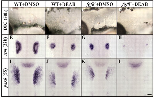
Fgf8 mediates otic induction in the absence of RA signaling. (A-D) Assessed by morphology at 50 hpf (DIC microscopy), otic vesicles are reduced after the depletion of RA (B) and in fgf8 mutants (C), and are completely lost in fgf8 mutants depleted of RA signaling (D), in comparison to controls (A). (E-H) Labeling with stm at 22 hpf reveals the presence of only residual otic cells in fgf8 mutants depleted of RA signaling (H) compared to fgf8 mutant (G), RA-depleted (F) or control (E) embryos, in which more otic cells are present. (I-L) Otic vesicle size reduction in fgf8 mutants depleted of RA signaling is evident as early as the preplacodal stages (five-somite stage, 5S), as detected by labeling with pax8. (A-D) Lateral views of live otic vesicles with anterior to the left and dorsal towards the top. (D-L) Dorsal views with anterior towards the top. Scale bar: 35 µm.
|