FIGURE

Fig. 4

ID
ZDB-FIG-070620-42
Publication
Hans et al., 2007 - Changes in retinoic acid signaling alter otic patterning
Other Figures
All Figure Page
Back to All Figure Page
Fig. 4

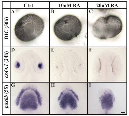
Increased RA signaling compromises lens specification. (A-F) Assessed both by morphology at 50 hpf (DIC microscopy, A-C) and cx44.1 expression at 24 hpf (D-F), the lens is reduced in size or completely lost after the application of 10 or 20 nM RA, compared with wild-type embryos. (G-I) Compromised lens specification is already evident at preplacodal stages (five-somite stage, 5S), as indicated by pax6b labeling. (A-C) Lateral views of live eyes with anterior to the left and dorsal towards the top. (D-I) Dorsal views with anterior towards the top. Scale bar: 30 µm for A-C; 50 µm for D-F; 35 µm for G-I.

Expression Data
Genes:
Fish:
Condition:
Anatomical Terms:
Stage Range: 5-9 somites to Prim-5

Expression Detail
Antibody Labeling
Phenotype Data

Phenotype Detail
Acknowledgments
This image is the copyrighted work of the attributed author or publisher, and ZFIN has permission only to display this image to its users. Additional permissions should be obtained from the applicable author or publisher of the image. Full text @ Development