|
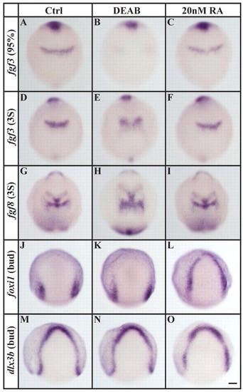
Loss and excess of RA signaling affect different tissues. (A-F) In comparison to controls (A,D) or to embryos treated with 20 nM RA (C,F), r4-specific fgf3 expression is delayed by the end of gastrulation in embryos depleted of RA signaling (DEAB, B) and remains reduced in an enlarged r4 primordium at the three-somite stage (E). (G-I) An enlarged r4-specific fgf8 expression domain is also present in embryos depleted of RA signaling (H) compared with control (G) or 20 nM RA-treated (I) embryos. (J-L) foxi1 expression is indistinguishable from control embryos (J) after RA-signaling depletion (K), whereas the application of 20 nM RA leads to ectopic expression within the entire preplacodal domain (L). (M-O) Expression of dlx3b in the preplacodal domain is unaffected by loss (N) or excess (O) of RA signaling. Dorsal views with anterior towards the top. Scale bar: 90 µm.
|