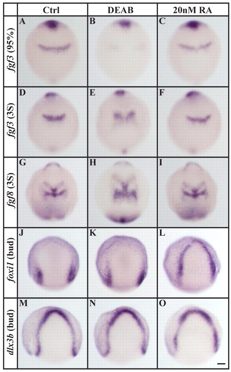
Fig. 3 Loss and excess of RA signaling affect different tissues. (A-F) In comparison to controls (A,D) or to embryos treated with 20 nM RA (C,F), r4-specific fgf3 expression is delayed by the end of gastrulation in embryos depleted of RA signaling (DEAB, B) and remains reduced in an enlarged r4 primordium at the three-somite stage (E). (G-I) An enlarged r4-specific fgf8 expression domain is also present in embryos depleted of RA signaling (H) compared with control (G) or 20 nM RA-treated (I) embryos. (J-L) foxi1 expression is indistinguishable from control embryos (J) after RA-signaling depletion (K), whereas the application of 20 nM RA leads to ectopic expression within the entire preplacodal domain (L). (M-O) Expression of dlx3b in the preplacodal domain is unaffected by loss (N) or excess (O) of RA signaling. Dorsal views with anterior towards the top. Scale bar: 90 µm.
Image
Figure Caption
Figure Data
Acknowledgments
This image is the copyrighted work of the attributed author or publisher, and
ZFIN has permission only to display this image to its users.
Additional permissions should be obtained from the applicable author or publisher of the image.
Full text @ Development

