
Fig. 4
- ID
- ZDB-FIG-061201-19
- Publication
- Shepherd et al., 2004 - Roles for GFRalpha1 receptors in zebrafish enteric nervous system development
- Other Figures
- All Figure Page
- Back to All Figure Page
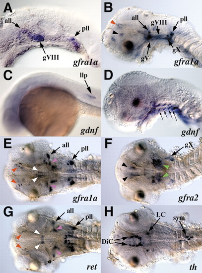
Expression of gdnf receptor components in cranial ganglia and brain. Lateral views (A-D) and dorsal views (E-H) of 24 hpf (A,C) and 48 hpf (B,D-H) embryos. At 24 hpf, gfra1a (A) is expressed in lateral line ganglia, while gdnf (C) is expressed in the migrating lateral line primordium (arrow). At 48 hpf, gfra1a (B) is expressed in epibranchial ganglia, while gdnf (D) is expressed in pharyngeal pouches (arrows). gfra1a (E), gfra2 (F), ret (G) and th (H) are shown at 48hpf. Black arrowheads indicate the ventral diencephalic neurons, red arrowheads indicate dorsal diencephalic neurons, white arrowheads indicate the ventral midbrain neurons, pink arrowheads indicate trigeminal motor nucleus, green arrowheads indicate anterior hindbrain domain. all, anterior lateral line ganglia; pll, posterior lateral line ganglia; llp, lateral line primordium; gV, trigeminal ganglia; gVI, facial ganglion; gVIII, octaval/statoacoustic ganglion; gIX, glossopharyngeal; gX, vagal ganglion; DiC, diencephalic catecholaminergic cell cluster; LC, locus coeruleus; sym, sympathetic neurons. |