|
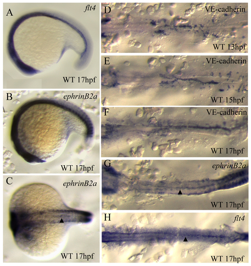
Expression of arterial and venous specific markers at the time of differentiation. Micrographs of in situ hybridization with flt4 (A,H), ephrinB2a (B,C,G) and VE-cadherin (cdh5) (D-F). Embryos at 13 hpf (A,D), 15 hpf (B,E) and 17 hpf (C,F), in lateral (A-B) and dorsal views (C-H; flat-mounted in D-H). Arrowheads in C,G,H indicate vascular expression of ephrinB2a (C,G), and flt4 (H). Both ephrinB2a and flt4 are expressed in the angioblasts located at the midline, but not those in the lateral plate mesoderm. Detailed expression patterns of arterial and venous endothelial cell markers can be found at the ZFIN website (www.zfin.org).
|