
FIGURE
Fig. 7
Fig. 7
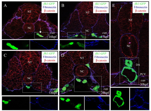
Angioblast migration in endodermless embryos. Transverse sections of endodermless embryos Tg(flk1:EGFP)s843;cas mutants visualized for GFP (green), fibronectin (blue), and ß-catenin (red). The GFP (green) and fibronectin (blue) signals of the outlined areas are shown separately (A-E). The sections shown are at the level of the 6th (A,B), 10th (C), 14th (D) and 18th (E) somites. White arrow in A shows a cluster of angioblasts exiting the LPM. Despite the absence of endoderm, angioblasts migrate to the midline. NT, neural tube; NC, notochord; DA, dorsal aorta; PCV, posterior cardinal vein. |
Expression Data
Expression Detail
Antibody Labeling
Phenotype Data
Phenotype Detail
Acknowledgments
This image is the copyrighted work of the attributed author or publisher, and
ZFIN has permission only to display this image to its users.
Additional permissions should be obtained from the applicable author or publisher of the image.
Full text @ Development