
- Title
-
Characterization of three novel members of the zebrafish Pax2/5/8 family: dependency of Pax5 and Pax8 expression on the Pax2.1 (noi) function
- Authors
- Pfeffer, P.L., Gerster, T., Lun, K., Brand, M., and Busslinger, M.
- Source
- Full text @ Development
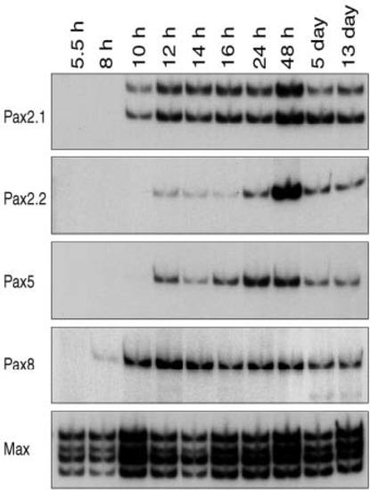
Temporal expression of the Pax2/5/8 genes during zebrafish embryogenesis. Total RNA of the indicated developmental stages was analyzed by quantitative RT-PCR using gene-specific primers. All cDNA sequences were amplified across the exon 5-exon 6 junction. The two PCR products of the Pax2.1 mRNA differ by the presence (upper band) or absence (lower band) of exon 5.1. The ubiquitiously expressed mRNA of the max gene (Kelly et al., 1995) was analyzed to quantitate the reverse-transcribed cDNA input of the PCR reaction. EXPRESSION / LABELING:
|
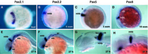
Expression of Pax2/5/8 genes at the midbrain-hindbrain boundary of zebrafish embryos. Transcripts of Pax2.1 (A,E), Pax2.2 (B,F), Pax5 (C,G) and Pax8 (D,H) were detected by whole-mount in situ hybridization in embryos from 10 hours (bud stage) to 27 hours postfertilization. Embryos are shown in lateral view (B-H) except for the bud stage (A) which is a dorsal view. cb, cerebellum; h, hindbrain; mhb, midbrainhindbrain boundary; na, (pro)nephric anlage; nd, nephric duct; pn, pronephros; os, optic stalk; otp, otic placode; otv, otic vesicle; ov, optic vesicle; ret, retina; som, somite; sc, spinal cord; tec, tectum; tg, tegmentum. |
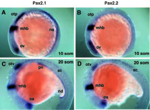
Comparison of the Pax2.1 and Pax2.2 expression patterns. The expression of Pax2.1 (A,C) and Pax2.2 (B,D) was analyzed by whole-mount in situ hybridization in 10- and 20-somite embryos (shown in lateral view). For abbreviations see legend to Fig. 3. EXPRESSION / LABELING:
|
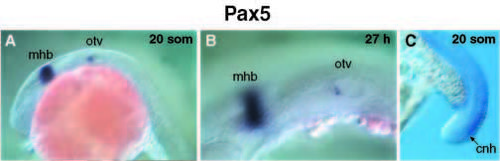
Pax5 expression in the otic vesicle and chordoneural hinge region. Whole-mount in situ hybridization of 20-somite (A) and 27- hour (B) embryos identified Pax5-expressing cells in the inner cell layer of the anterior otic vesicle (otv). At the 20-somite stage, weak Pax5 staining is also seen in the chordoneural hinge (cnh) region of the tail (C). EXPRESSION / LABELING:
|

Identification of novel expression domains of the vertebrate Pax8 gene. (A) Early Pax8 expression in the intermediate mesoderm (im) and in the anlage of the otic placode (otp). (B) Dorsal view of a 5-somite embryo with lateral expression of Pax8 in the otic placode and pronephric anlage (na). (C) Optical cross-section of the 5-somite embryo at the level of the otic placode (as indicated in B), which localizes the Pax8 staining in the ectoderm adjacent to the neural keel (nk). Lateral view (D) and higher magnifications (E,F) of a 20- somite embryo. Pax8 expression is no longer observed in the otic vesicle (otv), while it is maintained in the pronephros (out of focal plane) and nephric duct (nd). Arrows in E indicate individual Pax8- expressing neurons in the hindbrain, and numbers refer to individual rhombomeres. (G) Dorsal view of an 8.0-day mouse embryo (before somitogenesis) hybridized with a mouse Pax8 RNA probe. (H) Cross-section through the same embryo at the level indicated in G. Pax8 staining is detected lateral to the neuroepithelium (ne) of rhombomere B (rB) in the ectodermal layer corresponding to the otic placode. (I) Mouse Pax8 expression at the 12-somite stage. ha, hyoid arch; nc, notocord; ov, optic vesicle. EXPRESSION / LABELING:
|
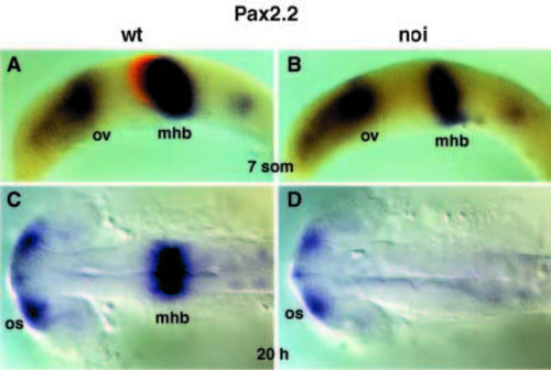
Pax2.2 expression is independent of the noi (Pax2.1) function. Transcripts of the Pax2.2 gene were analysed by whole-mount in situ hybridization in wild-type (A,C) and noitu29a mutant (B,D) embryos at 7 somites and 20 hours postfertilization. The 7-somite embryos (A,B; lateral view) were genotyped by double staining with Pax2.2 (blue) and eng3 (red) RNA probes. Homozygous noi mutant embryos were identified by the absence of engrailed (eng) expression at the midbrain-hindbrain boundary (mhb) (Brand et al., 1996). At 20 hours, the mhb tissue is entirely lost in noi mutant embryos (dorsal view). EXPRESSION / LABELING:
PHENOTYPE:
|
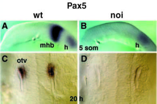
Dependency of Pax5 expression on the noi (Pax2.1) function. Pax5 expression was analyzed in wild-type (A,C) and noitu29a mutant (B,D) embryos at the 5-somite stage (lateral view) and 20 hours postfertilization (enlarged dorsal view, anterior to the top). For abbreviations see legend to Fig. 3. EXPRESSION / LABELING:
PHENOTYPE:
|
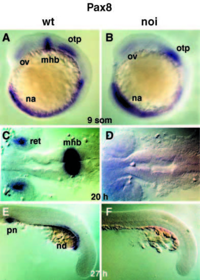
Requirement of the noi (Pax2.1) function for Pax8 expression. Wild-type (A,C,E) and noitu29a mutant (B,D,F) embryos were analyzed for Pax8 expression at the 9-somite stage (lateral view) as well as at 20 hours (dorsal view) and 27 hours postfertilization (lateral view). For abbreviations see legend to Fig. 3. EXPRESSION / LABELING:
PHENOTYPE:
|