
Image
|
Figure Caption
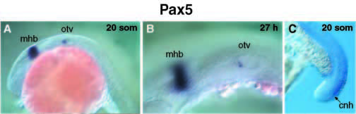
Fig. 5 Pax5 expression in the otic vesicle and chordoneural hinge region. Whole-mount in situ hybridization of 20-somite (A) and 27- hour (B) embryos identified Pax5-expressing cells in the inner cell layer of the anterior otic vesicle (otv). At the 20-somite stage, weak Pax5 staining is also seen in the chordoneural hinge (cnh) region of the tail (C).
Figure Data
Acknowledgments
This image is the copyrighted work of the attributed author or publisher, and
ZFIN has permission only to display this image to its users.
Additional permissions should be obtained from the applicable author or publisher of the image.
Full text @ Development