FIGURE SUMMARY
Title

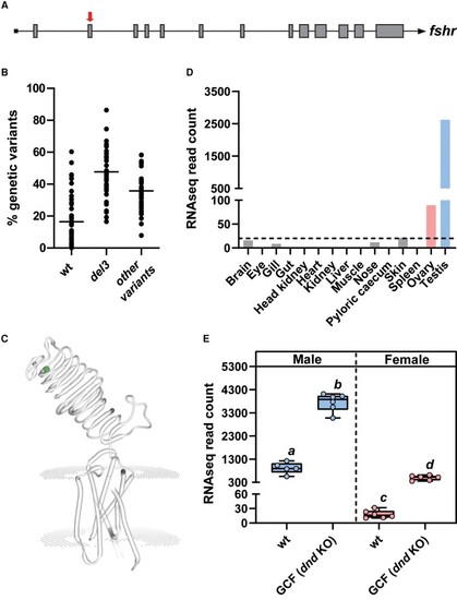
Loss of Fshr prevents testicular maturation in Atlantic salmon (Salmo salar L.)

Authors
Andersson, E., Schulz, R.W., Almeida, F., Kleppe, L., Skaftnesmo, K.O., Kjærner-Semb, E., Crespo, D., Fjelldal, P.G., Hansen, T.J., Norberg, B., Edvardsen, R.B., Wargelius, A.
Source
Full text @ Endocrinology

Fshr gene exon 2 target site (A; red arrow), The distribution of fshr variants in fin clips obtained from 40 fshr crispant males (B), dots representing prevalence of variant types found in single fish and the horizontal line the average percentage of each fshr variant groups which includes the wt fshr variant, deletion 3 (del3) variant and other fshr variants (mainly frameshift) deviating from wt fshr. In the SWISS-MODEL illustration of the Fshr protein (C), the CRISPR target site is indicated by the green dot. (D) fshr expression levels in juvenile tissues used for generating the reference annotation of the Atlantic salmon genome (GenBank GBRB00000000.1). Results are expressed as RNA sequencing (RNA-seq) normalized read counts (n = 1). Dashed line indicates 20 RNAseq reads. (E) Gonadal fshr expression in adult wild-type (wt) and germ cell–free (GCF) dead end (dnd) knockout salmon males and females. Data are shown as mean RNA-seq normalized read counts ± SEM (n = 5-6; different letters denote statistically significant differences).

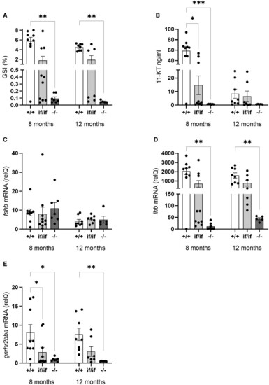
Gonadosomatic indices (GSI; A), plasma 11-ketotestosterone (11-KT; B), pituitary fshb (C), lhb (D), and gnrhr2bba (E) mRNA levels in F1 salmon males, showing fshr wild-type (+/+), double allelic in-frame (if/if), or double allelic loss of function mutation (−/−) genotypes, sampled at 8 and 12 months after the start of the maturation-inducing treatment (6 weeks of constant light and 16 °c water temperature, starting February 4, 2020). Data are presented as mean ± SEM. Significant differences between genotypes are indicated by asterisks (*P < .05, **P < .001, ***P < .0001).

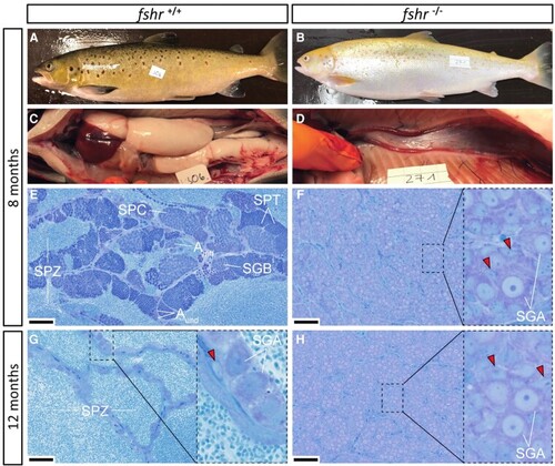
External appearance, macroscopic testis anatomy and histological sections of testes from mature wild-type control (fshr+/+) and F1 fshr−/− males sampled 8 months (A-F) and histological sections from testes sampled 12 months (G-H) after the start of the maturation-inducing treatment (6 weeks of constant light and 16 °C water temperature, starting February 4, 2020). At 8 months all but 1 control males showed the typical external appearance of maturing fish (A) and exhibited large testes upon dissection (C), containing all stages of spermatogenesis (E), including many spermatozoa in the tubular lumen. At 12 months, only 2 types of germ cells remained in the tubuli of control males: a large number of spermatozoa in the lumina and type A spermatogonia (SGA nuclei indicated) enveloped by 1 or 2 Sertoli cells (red arrowheads denote Sertoli cell nuclei) resided on the tubular wall (G). All F1 fshr−/− males showed an immature external appearance (B) and exhibited small testes (D) containing only type A spermatogonia (F, H). Bars indicate 50 µm. Aund, type A undifferentiated spermatogonia; Adiff, type A differentiating spermatogonia; SGB, type B spermatogonia; SPC, spermatocytes; SPT, spermatids; SPZ, spermatozoa.

Acknowledgments
This image is the copyrighted work of the attributed author or publisher, and ZFIN has permission only to display this image to its users. Additional permissions should be obtained from the applicable author or publisher of the image. Full text @ Endocrinology