FIGURE

Figure 3.

ID
ZDB-FIG-240221-32
Publication
Andersson et al., 2024 - Loss of Fshr prevents testicular maturation in Atlantic salmon (Salmo salar L.)
Other Figures
All Figure Page
Back to All Figure Page
Figure 3.

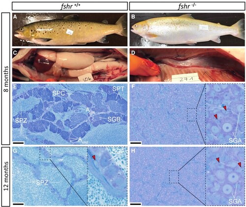
External appearance, macroscopic testis anatomy and histological sections of testes from mature wild-type control (fshr+/+) and F1 fshr−/− males sampled 8 months (A-F) and histological sections from testes sampled 12 months (G-H) after the start of the maturation-inducing treatment (6 weeks of constant light and 16 °C water temperature, starting February 4, 2020). At 8 months all but 1 control males showed the typical external appearance of maturing fish (A) and exhibited large testes upon dissection (C), containing all stages of spermatogenesis (E), including many spermatozoa in the tubular lumen. At 12 months, only 2 types of germ cells remained in the tubuli of control males: a large number of spermatozoa in the lumina and type A spermatogonia (SGA nuclei indicated) enveloped by 1 or 2 Sertoli cells (red arrowheads denote Sertoli cell nuclei) resided on the tubular wall (G). All F1 fshr−/− males showed an immature external appearance (B) and exhibited small testes (D) containing only type A spermatogonia (F, H). Bars indicate 50 µm. Aund, type A undifferentiated spermatogonia; Adiff, type A differentiating spermatogonia; SGB, type B spermatogonia; SPC, spermatocytes; SPT, spermatids; SPZ, spermatozoa.

Expression Data

Expression Detail
Antibody Labeling
Phenotype Data

Phenotype Detail
Acknowledgments
This image is the copyrighted work of the attributed author or publisher, and ZFIN has permission only to display this image to its users. Additional permissions should be obtained from the applicable author or publisher of the image. Full text @ Endocrinology