FIGURE SUMMARY
Title

A novel DSP zebrafish model reveals training- and drug-induced modulation of arrhythmogenic cardiomyopathy phenotypes

Authors
Celeghin, R., Risato, G., Beffagna, G., Cason, M., Bueno Marinas, M., Della Barbera, M., Facchinello, N., Giuliodori, A., Brañas Casas, R., Caichiolo, M., Vettori, A., Grisan, E., Rizzo, S., Dalla Valle, L., Argenton, F., Thiene, G., Tiso, N., Pilichou, K., Basso, C.
Source
Full text @ Cell Death Discov

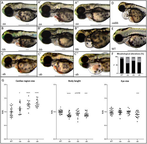
Cardiac alterations and developmental delay in Dsp mutant lines.

AF Mutant embryos (AC″, D) display cardiac alterations, compared to WT (E). Homozygous -aa (AA″) and -bb (BB″) mutant hearts appear dilated and/or structurally altered, with cardiac pericardial effusion and/or hemopericardium in the cardiac region. Double heterozygotes -ab (CC″) show a more serious phenotype than -aa and -bb lines. Double homozygotes -aabb display the most severe phenotype (D). F Percentage of heart alteration in different genotypes. Sample size: n = 100. G Cardiac size analysis shows that all mutants present heart dilation. Body length and eye size measurements indicate developmental delay in mutants, especially in the -ab condition. All embryos are at 3 dpf in lateral view, anterior to the left. Sample size Cardiac region size: WT n = 31; -aa n = 18; -bb n = 16; -ab n = 16. Sample size Body length: WT n = 32; -aa n = 22; -bb n = 19; -ab n = 21. Sample size Eye size: WT n = 32; -aa n = 22; -bb n = 19; -ab n = 21. R.R. relative ratio. Error bars: SEM. *p < 0.05; **p < 0.01; ***p < 0.001; ****p < 0.0001. Test: One-way ANOVA followed by Tukey’s test.

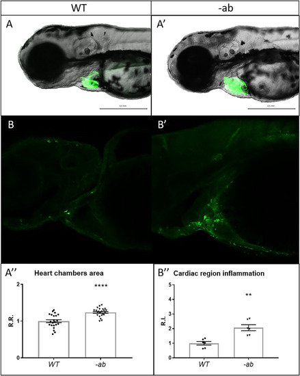
Detection of cardiac dilation and inflammation in zebrafish Dsp mutants.

AA′-A″ Heart morphology analysis of wild type and -ab mutant zebrafish embryos using a myocardial-specific transgene Tg(tg:EGFP-myl7:EGFP)ia300 identified a global dilation of both chambers in -ab mutants. All embryos are at 3 dpf, in lateral view. Sample size: n = 22 ± 1. R.R relative ratio. Error bars: SEM. ****p < 0.0001. Test: Unpaired t-test. BB′–B″: Cells expressing the inflammatory marker L-plastin are more abundant in -ab mutants, compared to WT. All embryos are at 3 dpf, in lateral view, anterior to the left. Sample size: n = 6. R.I. relative intensity. Error bars: SEM. **p < 0.01. Test: Unpaired t-test.

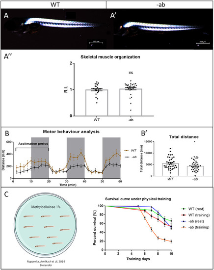
Impaired motor behaviour and exercise-induced mortality in zebrafish Dsp mutant larvae.

AA′–A″: Birefringence analysis on WT and mutant larvae at 5 dpf does not detect any significant difference in the skeletal muscles structure. All embryos are at 5 dpf, in lateral view, anterior to the left. Sample size: WT n = 20; -ab n = 30. R.I. relative intensity. Error bars: SEM. ns not significant. Test: Unpaired t-test. BB′: WT and -ab larvae at 5 dpf display normal response to light (white areas) and dark (grey areas) stimuli, but with reduced motor performances if mutated, evaluated as total distance swum (B). Sample size: n = 36. Error bars: SEM. *p < 0.05. Test: Unpaired t-test. C A mild training protocol was applied on larvae from 3 to 13 dpf, for 10 days. A statistically significant increase in mortality was observed in trained mutants when compared with resting controls (p < 0.0001). Sample size: n = 50. Error bars: SEM. Test: Log-rank (Mantel–Cox) test.

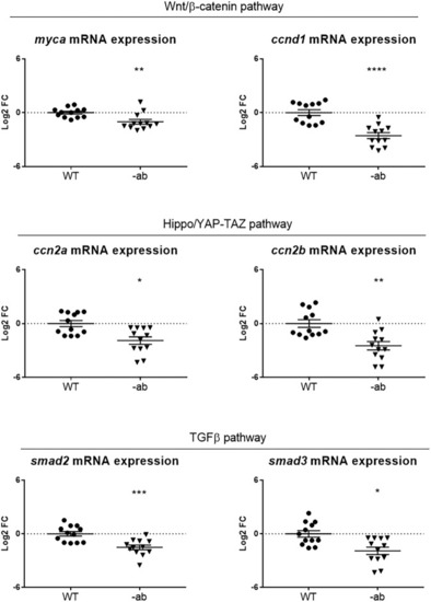
Signalling pathways dysregulation in Dsp mutant adult hearts.

qPCR analysis of the expression of Wnt/β-catenin, YAP-TAZ and TGF-β signalling members showed a downregulation of all pathways in 1-year old Dsp mutant hearts. Each point on the graph corresponds to a pool of 3 hearts of the same genotype. Sample size: n = 12. Log2 FC: Log2 Fold Change. Error bars: SEM. *p < 0.05; **p < 0.01; ***p < 0.001; ****p < 0.0001. Test: Unpaired t-test.

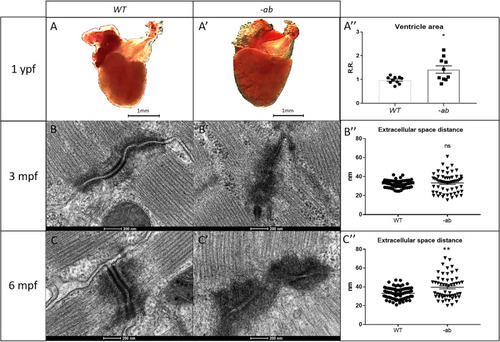
Cardiac dilation and TEM analysis of 3- and 6-month old Dsp mutant zebrafish hearts.

AA′-A″: 1-year old -ab mutant ventricles showed a statistically significantly dilation (P < 0.05) in comparison with WT controls. Sample size: n = 10. Error bars: SEM. R.R relative ratio. *p < 0.05. Test: Unpaired t-test. BB′–B″–CC′–C″: TEM analysis of 3- and 6-month-old mutated zebrafish heart revealed “pale”, disorganized and delocalized desmosomes. 6-month old -ab hearts showed a significantly increased distance in the extracellular space. Sample size: n = 60 ± 10 desmosome in 3 mutated and WT hearts. Error bars: SEM. ns not significant; **p < 0.01. Test: Unpaired t-test.

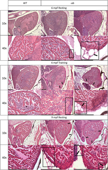
Cardiac dilation and structural changes in Dsp mutant hearts

Histological analysis of 6-month old -ab mutant zebrafish showed mild rarefaction of cardiomyocytes, thinning of the myocardial layer, age-related alterations in the distribution and organization of the trabeculae network, an abnormal shape of the ventricle, the presence of possible vessels dilation (rectangular box) and accumulation of adipose cells outside and inside the myocardial layer, in ≈50% of analyzed fish (square boxes and black arrows). Histological analysis of 6-month old mutated zebrafish confirmed the worsening of the condition after intensive physical training, like vessels dilation (rectangular box) and a more intrusive presence of adipose cells, in ≈80% of analyzed fish (square boxes and black arrows), showing similarities with 9-month old mutant hearts at rest. 9-month-old -ab mutant zebrafish showed worsening of the cardiac phenotype compared to 6-month old mutant hearts, with thickness of the myocardial layer, vessels dilation (rectangular box), and more intrusive presence of adipose cells, in ≈80% of analyzed fish (square boxes and black arrows). Sample size: n = 3 for each condition and age. Scale bar: 200 μm.

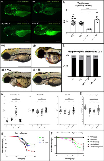
Wnt/β-catenin activation rescues AC phenotypes in Dsp zebrafish mutants.

A -ab mutant zebrafish larvae in Wnt/β-catenin Tg(7xTCF-Xla.Siam:EGFP)ia4 transgenic background, treated for 2 days with 5 µM XAV939 (XAV, Wnt inhibitor) or 40 µM SB216763 (SB, Wnt agonist), showed a downregulation or a full recovery of the pathway activity (GFP reporter) when compared to WT controls or untreated mutants. All embryos are at 3 dpf and displayed in lateral view, anterior to the left. Sample size: WT n = 45; -ab n = 55; -ab + XAV n = 17; -ab + SB n = 21. R.I. relative intensity. Error bars: SEM. **p < 0.01; ****p < 0.0001. Test: One-way ANOVA followed by Tukey’s test. B The -ab larvae treated with 40 µM SB showed a significant reduction of cardiac dysmorphisms, while 5 µM XAV induced a worsening of the condition when compared to WT controls or untreated mutants. All embryos are at 3 dpf and displayed in lateral view, anterior to the left. Sample size: n = 100. C The -ab larvae treated for 2 days with 40 µM SB216763 showed a significant rescue of the main morphological alterations. The treatment with 5 µM XAV939 induced a worsening of the condition. Sample size Cardiac region size: WT n = 41; -ab n = 41; -ab + XAV n = 37; -ab + SB n = 82. Sample size Body length WT n = 41; -ab n = 46; -ab + XAV n = 54; -ab + SB n = 82. Sample size Eye size: WT n = 41; -ab n = 46; -ab + XAV n = 36; -ab + SB n = 81. R.R. relative ratio. Error bars: SEM. ns not significant; **p < 0.01; ***p < 0.001; ****p < 0.0001. Test: One-way ANOVA followed by Tukey’s test. D At 5 dpf, treated -ab larvae showed a statistically significant recovery of the bradycardia phenotype. Sample size: WT n = 70; -ab n = 66; -ab + XAV n = 40; -ab + SB n = 77. Error bars: SEM. ****p < 0.0001. Test: One-way ANOVA followed by Tukey’s test. E One-month survival rate of WT and -ab larvae untreated or treated with 40 µM SB216763 for 4 days; a statistically significant decreased mortality in mutants is observed after treatment, compared to untreated mutants (p < 0.0001). Sample size: n = 100. Error bars: SEM. Test: Log-rank (Mantel-Cox) test. F: 5-day survival analysis, from 3 to 8 dpf, of untrained or trained WT and -ab, as well as -ab simultaneously SB-treated and trained (training induced in 1% methylcellulose). Trained -ab, treated with SB, showed a statistically significant decrease in mortality when compared with -ab subjected to training in the absence of SB (p < 0.01). Sample size: n = 50. Error bars: SEM. Test: Test: Log-rank (Mantel–Cox) test.

Acknowledgments
This image is the copyrighted work of the attributed author or publisher, and ZFIN has permission only to display this image to its users. Additional permissions should be obtained from the applicable author or publisher of the image. Full text @ Cell Death Discov