IMAGE

Fig. 2

ID
ZDB-IMAGE-231208-30
Source
Figures for Celeghin et al., 2023
Image
Figure Caption

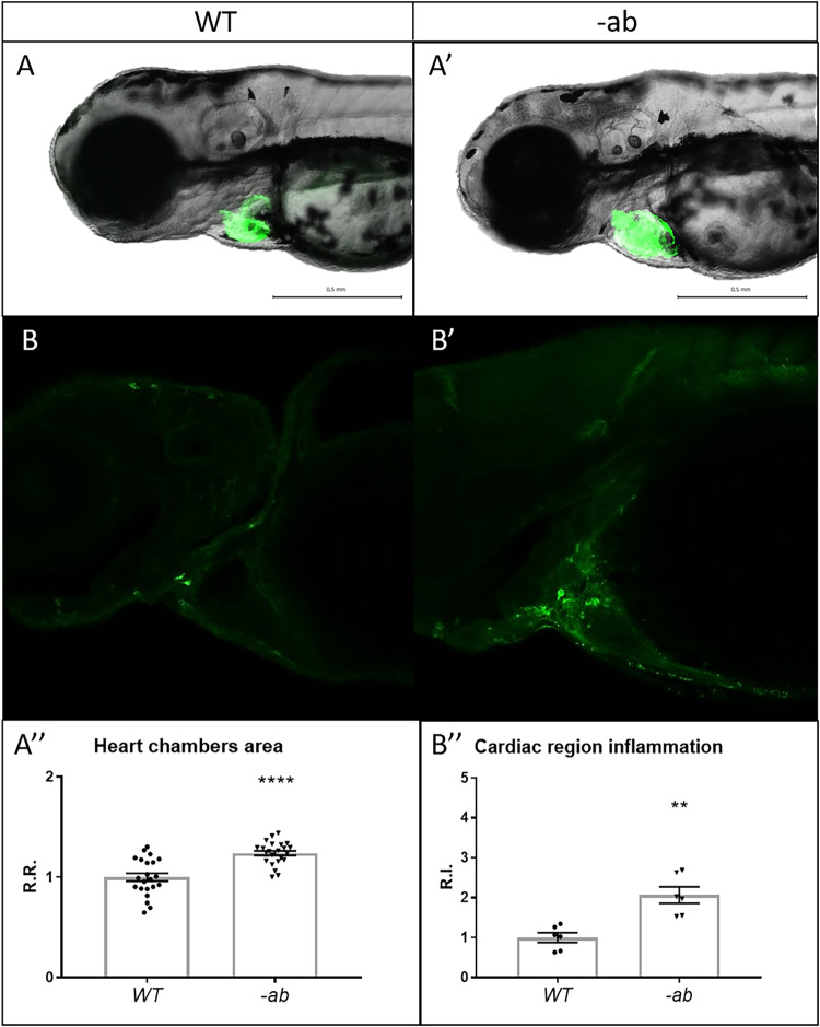
Fig. 2 Detection of cardiac dilation and inflammation in zebrafish Dsp mutants.

AA′-A″ Heart morphology analysis of wild type and -ab mutant zebrafish embryos using a myocardial-specific transgene Tg(tg:EGFP-myl7:EGFP)ia300 identified a global dilation of both chambers in -ab mutants. All embryos are at 3 dpf, in lateral view. Sample size: n = 22 ± 1. R.R relative ratio. Error bars: SEM. ****p < 0.0001. Test: Unpaired t-test. BB′–B″: Cells expressing the inflammatory marker L-plastin are more abundant in -ab mutants, compared to WT. All embryos are at 3 dpf, in lateral view, anterior to the left. Sample size: n = 6. R.I. relative intensity. Error bars: SEM. **p < 0.01. Test: Unpaired t-test.

Acknowledgments
This image is the copyrighted work of the attributed author or publisher, and ZFIN has permission only to display this image to its users. Additional permissions should be obtained from the applicable author or publisher of the image. Full text @ Cell Death Discov