
Fig. 6
- ID
- ZDB-FIG-231208-32
- Publication
- Celeghin et al., 2023 - A novel DSP zebrafish model reveals training- and drug-induced modulation of arrhythmogenic cardiomyopathy phenotypes
- Other Figures
- All Figure Page
- Back to All Figure Page
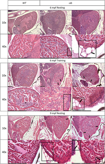
Cardiac dilation and structural changes in Dsp mutant hearts Histological analysis of 6-month old -ab mutant zebrafish showed mild rarefaction of cardiomyocytes, thinning of the myocardial layer, age-related alterations in the distribution and organization of the trabeculae network, an abnormal shape of the ventricle, the presence of possible vessels dilation (rectangular box) and accumulation of adipose cells outside and inside the myocardial layer, in ≈50% of analyzed fish (square boxes and black arrows). Histological analysis of 6-month old mutated zebrafish confirmed the worsening of the condition after intensive physical training, like vessels dilation (rectangular box) and a more intrusive presence of adipose cells, in ≈80% of analyzed fish (square boxes and black arrows), showing similarities with 9-month old mutant hearts at rest. 9-month-old -ab mutant zebrafish showed worsening of the cardiac phenotype compared to 6-month old mutant hearts, with thickness of the myocardial layer, vessels dilation (rectangular box), and more intrusive presence of adipose cells, in ≈80% of analyzed fish (square boxes and black arrows). Sample size: |
| Fish: | |
|---|---|
| Condition: | |
| Observed In: | |
| Stage: | Adult |