IMAGE

Fig. 5

ID
ZDB-IMAGE-231208-33
Source
Figures for Celeghin et al., 2023
Image
Figure Caption

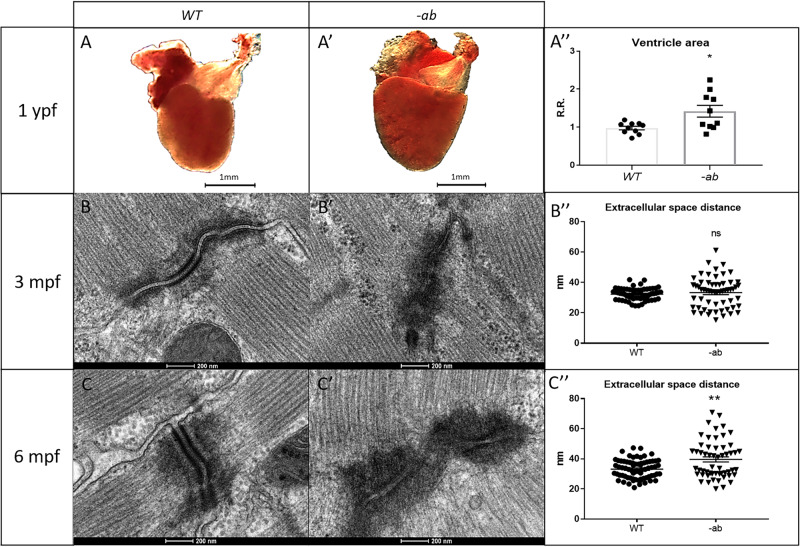
Fig. 5 Cardiac dilation and TEM analysis of 3- and 6-month old Dsp mutant zebrafish hearts.

AA′-A″: 1-year old -ab mutant ventricles showed a statistically significantly dilation (P < 0.05) in comparison with WT controls. Sample size: n = 10. Error bars: SEM. R.R relative ratio. *p < 0.05. Test: Unpaired t-test. BB′–B″–CC′–C″: TEM analysis of 3- and 6-month-old mutated zebrafish heart revealed “pale”, disorganized and delocalized desmosomes. 6-month old -ab hearts showed a significantly increased distance in the extracellular space. Sample size: n = 60 ± 10 desmosome in 3 mutated and WT hearts. Error bars: SEM. ns not significant; **p < 0.01. Test: Unpaired t-test.

Acknowledgments
This image is the copyrighted work of the attributed author or publisher, and ZFIN has permission only to display this image to its users. Additional permissions should be obtained from the applicable author or publisher of the image. Full text @ Cell Death Discov