
- Title
-
Reversibility of thyroid hormone system-disrupting effects on eye and thyroid follicle development in zebrafish (Danio rerio) embryos
- Authors
- Pannetier, P., Poulsen, R., Gölz, L., Coordes, S., Stegeman, H., Koegst, J., Reger, L., Braunbeck, T., Hansen, M., Baumann, L.
- Source
- Full text @ Environ. Toxicol. Chem.
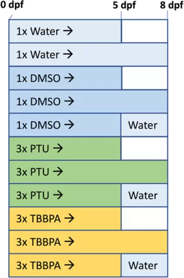
Experimental design of exposure experiments with zebrafish. For each control and exposure concentration, three conditions were run: exposure from 0 to 5 days postfertilization (dpf), exposure from 0 to 8 dpf, and exposure from 0 to 5 dpf with subsequent recovery in water until 8 dpf. Three independent replicate runs with three test concentrations of propylthiouracil or tetrabromobisphenol A plus water and solvent control were performed separately for each endpoint. For histopathology, 24 embryos per replicate run and concentration were used. For thyroid follicle analyses, 20 embryos per replicate run and concentration were used. For thyroid hormone measurements, 40 embryos per replicate run and concentration were used. DMSO = dimethyl sulfoxide; PTU = propylthiouracil; TBBPA = tetrabromobisphenol A. |
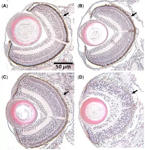
Semiquantitative severity grading of the loss of pigmentation in the retinal pigment epithelium (indicated by arrows): To account for the different intensity levels of pigmentation loss, four grades were assigned: (A) no effect (control), (B) weak effect (low propylthiouracil [PTU] concentration), (C) clear effect (medium PTU concentration), (D) strong effect (high PTU concentration), after 5 days of exposure. |
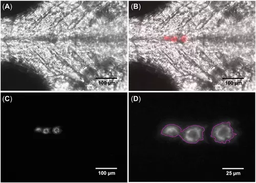
Fiji (ImageJ Ver 2.1.0/1.53c) analysis of the thyroid follicles of a negative control transgenic zebrafish (Danio rerio, tg:mcherry) embryo at ×20 magnification: (A) brightfield (DIA 340 mm) image of the head region; (B) overlay of the DIA and tetramethylrhodamine (TRITC) channel showing the location of thyroid follicles in red; (C) TRITC channel as gray scale used for the image analysis; and (D) thyroid follicle micrograph after image analysis. Three pictures with average intensity were overlaid, and the area within the pink circles was analyzed (D). |
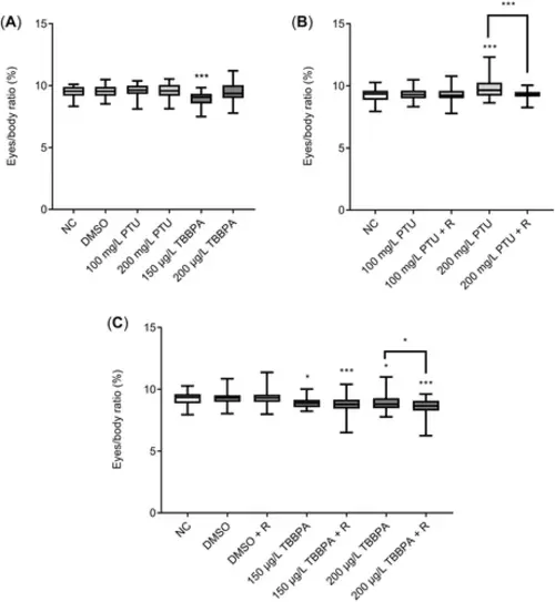
Relative eye size of zebrafish (Danio rerio) embryos and larvae after 5 (A) and 8 days after exposure with or without recovery phase to propylthiouracil (B) and tetrabromobisphenol A (C). Data given as means ± SEM for n = 3 replicates (10–29 individuals). Because of technical issues, data from one replicate were not analyzed. Statistically significant differences from the negative control or between continued exposure and recovery groups: *p < 0.05, **p < 0.01, ***p < 0.001. NC = negative control (water); DMSO = dimethyl sulfoxide (solvent control, 0.002%); PTU = propylthiouracil; TBBPA = tetrabromobisphenol A; R = recovery (3 days). |
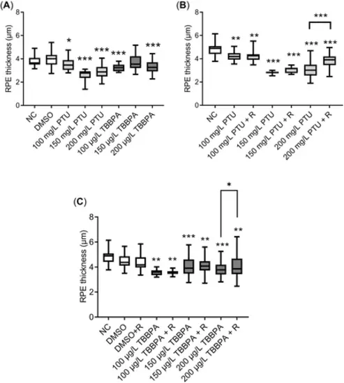
Retinal pigment epithelium thickness of zebrafish larvae at 5 days postfertilization (dpf; A) and 8 dpf after exposure with or without recovery phase to propylthiouracil (B) and tetrabromobisphenol A (C). Data given as means ± SEM for n = 4 replicates (10–15 individuals). Statistically significant differences from the negative control or between continued exposure and recovery groups: *p < 0.05, **p < 0.01, ***p < 0.001. RPE = retinal pigment epithelium; NC = negative control (water); DMSO = dimethyl sulfoxide (solvent control, 0.002%); PTU = propylthiouracil; TBBPA = tetrabromobisphenol A; R = recovery (5-day exposure with the respective concentration and a 3-day recovery in clean water). |
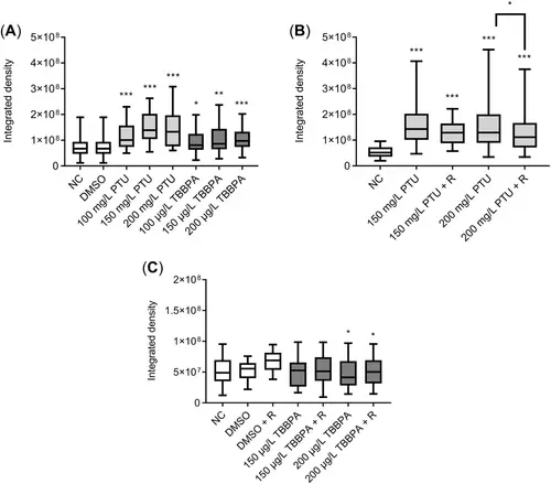
Integrated density of thyroid follicles of transgenic zebrafish larvae at 5 days postfertilization (dpf; A) and 8 dpf after exposure with or without recovery phase to propylthiouracil (B) and tetrabromobisphenol A (C). Data given as means ± SEM for n = 4 replicates (10–15 individuals per replicate). Statistically significant differences from the negative control or between continued exposure and recovery groups: *p < 0.05, **p < 0.01, ***p < 0.001. NC = negative control (water); DMSO = dimethyl sulfoxide (solvent control, 0.002%); PTU = propylthiouracil; TBBPA = tetrabromobisphenol A; R = recovery (5-day exposure with the respective concentration and a 3-day recovery in clean water). |
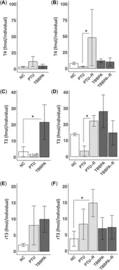
Thyroid hormones thyroxine (A,B), triiodothyronine (C,D), and triiodothyronine (E,F) in whole-body samples at 5 days postfertilization (dpf; A,C,E) and 8 dpf (B,D,F). Propylthiouracil and tetrabromobisphenol A concentrations were 200 mg/L and 200 µg/L, respectively. Data given as means ± SEM for n = 3 replicates (20 individuals per replicate). Statistically significant difference: *p < 0.05. T4 = thyroxine; <LoD = all replicates were below the limit of detection (LoD), and a replacement value equal to one half the LoD was used; NC = negative control (water); PTU = propylthiouracil; TBBPA = tetrabromobisphenol A; R = recovery (5-day exposure with the respective concentration and a 3-day recovery in water); T3 = triiodothyronine; rT3 = reverse-triiodothyronine. |
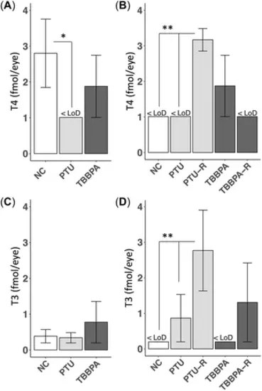
Thyroid hormones thyroxine (A,B) and triiodothyronine (C,D) in eyes at 5 days postfertilization (dpf; A,C) and 8 dpf (B,D). Propylthiouracil and tetrabromobisphenol A concentrations were 200 mg/L and 200 µg/L, respectively. Data given as means ± SEM for n = 3 replicates (40 eyes per replicate). Statistically significant differences: *p < 0.05, **p < 0.01. T4 = thyroxine; <LoD = all replicates were below the limit of detection (LoD) and a replacement value equal to one half the LoD was used; NC = negative control (water); PTU = propylthiouracil; TBBPA = tetrabromobisphenol A; R = recovery (5-day exposure with the respective concentration and a 3-day recovery in artificial water); T3 = triiodothyronine. |