FIGURE SUMMARY
Title

Control of Directed Cell Migration after Tubular Cell Injury by Nucleotide Signaling

Authors
Gessler, S., Guthmann, C., Schuler, V., Lilienkamp, M., Walz, G., Yakulov, T.A.
Source
Full text @ Int. J. Mol. Sci.

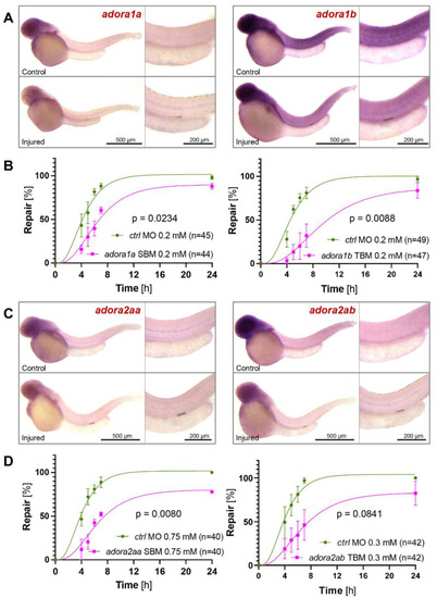
The involvement of adenosine receptors in zebrafish pronephros repair. (A) In situ hybridization revealed upregulation of zebrafish adora1a and adora1b after a laser-induced injury. Images were obtained two hours after the injury. (B) Depletion of zebrafish adora1a or adora1b by splice- (SBM) or translation- (TBM) blocking morpholino oligonucleotides (MO) delayed the repair process. (C) In situ hybridization revealed upregulation of zebrafish adora2aa and adora2ab after a laser-induced injury. Images were obtained two hours after the injury. (D) Depletion of zebrafish adora2aa or adora2ab by SBM delayed the repair process (mean ± SEM; 2-way ANOVA).

The involvement of adenosine deaminase family members in zebrafish pronephros repair. (A) In situ hybridization revealed upregulation of zebrafish ada. Depletion of zebrafish ada by a splice—(SBM) blocking morpholino oligonucleotides (MO) delayed the repair process. (B) In situ hybridization revealed upregulation of zebrafish ada2a. Depletion of zebrafish ada2a by a translation- (TBM) blocking morpholino oligonucleotides (MO) had no effect on the repair process. (C) In situ hybridization revealed upregulation of zebrafish ada2b. Depletion of zebrafish ada2b by a TBM delayed the repair process. (D) In situ hybridization revealed upregulation of zebrafish adal. Depletion of zebrafish adal by a TBM had no significant influence on the repair process (mean ± SEM; 2-way ANOVA).

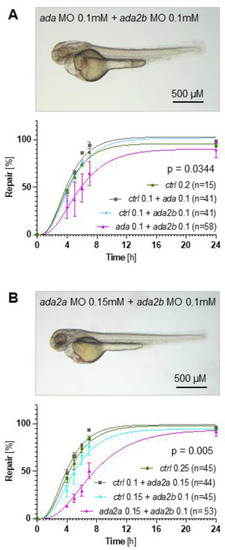
Synergistic effects between adenosine deaminase family members. (A) Combining low concentrations of the ada splice-blocking morpholino oligonucleotide (MO) and the ada2b translation-blocking MO did not affect zebrafish embryogenesis. However, the combination significantly delayed the repair process in comparison to the control (ctrl) or either single MO. (B) Combining low concentrations of the ada2a translation-blocking MO and the ada2b translation-blocking MO did not affect zebrafish embryogenesis. However, the combination significantly delayed the repair process in comparison to the control (ctrl) or either single MO (mean ± SEM; 2-way ANOVA).

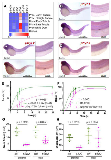
The involvement of purinergic P2ry2 receptors in zebrafish pronephros repair. (A) Single-cell RNA sequencing, performed with two-day-old zebrafish embryos, revealed expression of p2ry2 along the zebrafish embryo, while other P2 family members were expressed at low levels. (BD) In situ hybridization revealed upregulation of all three zebrafish p2ry2 variants two hours after a laser-induced injury. (E) Depletion of zebrafish p2ry2.1, p2ry2.2 and p2ry2.3 by translation- (TBM) blocking morpholino oligonucleotides (MO) (each MO, 0.2 mM) significantly delayed the repair process. (F) Depletion of zebrafish p2ry2.1, p2ry2.2 and p2ry2.3 by CRISPR/Cas9 in combination with 8 sgRNAs delayed the repair process; however, the difference was statistically not significant (mean ± SEM; 2-way Anova). (G,H) High-resolution video microscopy revealed that p2ry2 depletion reduced track speed and cell displacement particularly in the proximal parts of the pronephros (mean ± SD; Mann–Whitney test).

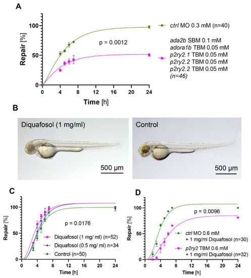
Effect of the combined adenosine pathway knockdown, and of the P2YR2 agonist Diquafosol on the repair process. (A) Combined depletion of ada2b, adora1b, p2ry2.1, p2ry2.2 and p2ry2.3 with low concentrations of MOs significantly delayed the repair process (mean ± SEM; 2-way Anova). (B) Diquafosol (1 mg/mL) did not affect zebrafish development. (C) Diquafosol exposure for 3 h before, and for 24 h after laser-induced injury resulted in accelerated repair (mean ± SEM; 2-way Anova). (D) Depletion of zebrafish p2ry2 variants by translation-blocking morpholino oligonucleotides (MO) prevented the accelerating effect of Diquafosol.

Proposed ATP-dependent signaling after a laser-induced zebrafish pronephros injury. Damaged cells release nucleotides, including ATP. Released ATP is metabolized by ectonucleoside triphosphate diphosphohydrolase-1 (ENTPD1, CD39) and ecto-5′-nucleotidase (NT5E, CD73) to adenosine. Adenosine is rapidly removed from the extracellular environment by equilibrative nucleoside transporters (ENTs), or metabolized to inosine by extracellular adenosine deaminase (ADA), associated with CD26 or adenosine receptors. Intracellular adenosine is metabolized by cytoplasmic ADA. The G protein-coupled P2RY2 receptor, signaling through Gi/o, activates PLCß, while the adenosine A2A receptor stimulated adenylyl cyclase (AC) and cAMP production through Gs. Note that other adenosine family members couple to Gi/o, inhibiting AC.

Acknowledgments
This image is the copyrighted work of the attributed author or publisher, and ZFIN has permission only to display this image to its users. Additional permissions should be obtained from the applicable author or publisher of the image. Full text @ Int. J. Mol. Sci.