
- Title
-
Identification of 4 novel human ocular coloboma genes ANK3, BMPR1B, PDGFRA, and CDH4 through evolutionary conserved vertebrate gene analysis
- Authors
- Owen, N., Toms, M., Young, R.M., Eintracht, J., Sarkar, H., Brooks, B.P., Moosajee, M., Genomics England Research Consortium
- Source
- Full text @ Genet. Med.
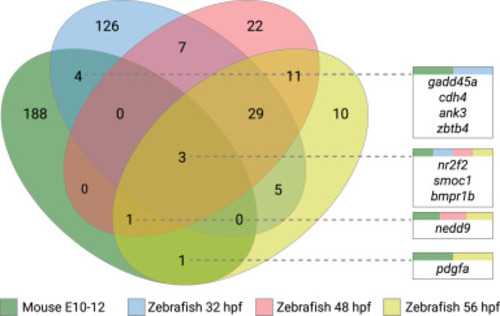
Figure 1. Venn diagram of the significantly regulated genes identified in mouse and zebrafish optic fissure developmental time points. The figure shows comparisons between differentially expressed gene lists of mouse and zebrafish optic fissure vs retinal tissue, with the numeric value indicating the number of genes at each intersection. Identified commonalities of genes are represented in each box, with colors representing each data set intersection. hpf, hours post fertilization. |
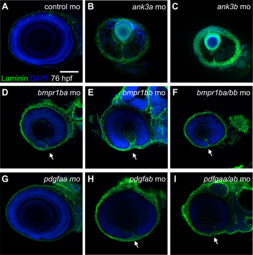
Figure 2. Knockdown analysis of ventral retina and optic fissure specific genes in zebrafish. Confocal imaging of lateral views (dorsal up, anterior left) of immunofluorescence staining of laminin protein in zebrafish embryos at 76 hours post fertilization in (A) control MO or MO knockdowns of (B) ank3a, (C) ank3b, (D) bmpr1ba, (E) bmpr1bb, (F) bmpr1ba/bmpr1bb, (G) pdgfaa, (H) pdgfab, and (I) pdgfaa/pdgfab mutant embryos. Scale bar = 100 μm. MO, morpholino. PHENOTYPE:
|
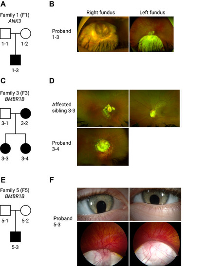
Figure 3. Pedigrees and clinical imaging of 3 unrelated coloboma families. A. F1 is a White British nonconsanguineous pedigree with 1 affected male patient (proband 1-3) harboring a de novo sporadic heterozygous missense variant in ANK3 NM_020987.5:c.11650A>T, p.(Thr3884Ser). B. Corresponding widefield color fundus photographs of the right and left eye of the proband aged 17 years showing bilateral inferior chorioretinal coloboma with extensive optic disc involvement (right eye is more severely affected than the left eye as indicated also by the residual best corrected visual acuity). C. F3 is an Indian nonconsanguineous pedigree with autosomal dominant inheritance; the mother and the 2 daughters were affected with a heterozygous missense variant in BMPR1B NM_001203.2:c.272G>T, p.(Arg91Ile). D. Widefield color fundus photograph of the right eye of the proband (3-4) aged 13 showed a large optic disc coloboma extending inferiorly with a region of associated retinal pigment epithelium atrophy below this. The left eye image was not available. Widefield color fundus photographs of the right and left eye of the proband (3-3) aged 18 showing bilateral optic disc coloboma sparing the macula, with irregular asymmetrical peripapillary atrophy in the right eye. E. F5 is a White Northern European nonconsanguineous pedigree with 1 affected male patient (proband 5-3) with a de novo sporadic heterozygous missense variant in BMPR1B NM_001203.2:c.671 G>A, p.(Arg224His). F. Anterior segment color photographs of the right and left eye of the proband aged 4 showing bilateral inferior iris coloboma and corresponding widefield color fundus photographs showing bilateral inferior chorioretinal coloboma with extensive optic disc and macular involvement. F1, family 1; F3, family 3; F5, family 5. |
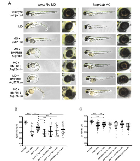
Figure 4. MO knockdown and rescue experiments of bmpr1ba/b in zebrafish. Injection of the MOs alone showed reduction in eye size and the presence of a coloboma. A. Coinjection with in vitro synthesized human BMPR1B messenger RNA (mRNA) rescues the phenotypic effects of the MO alone. Human variants identified within the Genomics England cohort were expressed and mRNAs coinjected with each MO. Scale bar = 500 μm. Eye diameter was reported for (B) bmpr1ba and (C) bmpr1bb compared with wild-type uninjected; MO; MO + human wild-type BMPR1B; and variants Arg91Ile, Arg224His, Arg224Leu, and Arg376Glu. (n for bmpr1ba experiments = 23, 25, 55, 13, 21, 28, 42 and n for bmpr1bb experiments = 23, 56, 55, 32, 34, 33, 24). Unpaired t tests were used to compare data. ∗∗P < .01, ∗∗∗P < .001, ∗∗∗∗P < .0001, ns P > .05. MO, morpholino; ns, nonsignificant. PHENOTYPE:
|

ZFIN is incorporating published figure images and captions as part of an ongoing project. Figures from some publications have not yet been curated, or are not available for display because of copyright restrictions. |
|
ZFIN is incorporating published figure images and captions as part of an ongoing project. Figures from some publications have not yet been curated, or are not available for display because of copyright restrictions. |