
Image
|
Figure Caption
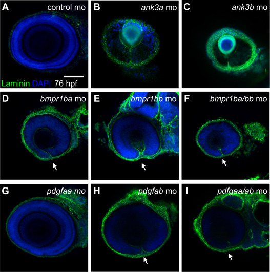
Fig. 2 Figure 2. Knockdown analysis of ventral retina and optic fissure specific genes in zebrafish. Confocal imaging of lateral views (dorsal up, anterior left) of immunofluorescence staining of laminin protein in zebrafish embryos at 76 hours post fertilization in (A) control MO or MO knockdowns of (B) ank3a, (C) ank3b, (D) bmpr1ba, (E) bmpr1bb, (F) bmpr1ba/bmpr1bb, (G) pdgfaa, (H) pdgfab, and (I) pdgfaa/pdgfab mutant embryos. Scale bar = 100 μm. MO, morpholino.
Figure Data
Acknowledgments
This image is the copyrighted work of the attributed author or publisher, and
ZFIN has permission only to display this image to its users.
Additional permissions should be obtained from the applicable author or publisher of the image.
Full text @ Genet. Med.