FIGURE SUMMARY
Title

An Inhibitor of NF-κB and an Agonist of AMPK: Network Prediction and Multi-Omics Integration to Derive Signaling Pathways for Acteoside Against Alzheimer's Disease

Authors
Li, Y.Q., Chen, Y., Jiang, S.Q., Shi, Y.Y., Jiang, X.L., Wu, S.S., Zhou, P., Wang, H.Y., Li, P., Li, F.
Source
Full text @ Front Cell Dev Biol

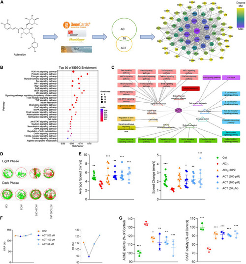
ACT attenuated AlCl3-induced AD in zebrafish larvae. (A) The workflow of ACT-AD targets interaction network modeling. The size and color of nodes represented the degree of targets. The width of edges was consistent with the combined score of targets. (B) Top 30 of KEGG enrichment. (C) Illustration of KEGG pathway enrichment analysis. (D) The representative swimming trails of zebrafish larvae in the light and dark phase. (E) The average speed and speed change in different groups (n = 10). (F) DRR (%) and RE (%) in the drug treatment groups (n = 10). (G) The evaluation of AChE and ChAT activities (n = 3). *P < 0.05, **P < 0.01, and ***P < 0.005, versus the AlCl3 group. DPZ as the positive control.

ACT regulated M1/M2 polarization in LPS-stimulated BV-2 cells. (A) Effects of ACT on LPS-induced BV-2 cell viability. (B) Cell morphology with IBA-1 staining was observed with inverted phase contrast fluorescence microscope (×10). The control group exhibited small soma with distal arborization, showing the typical ramified morphology of resting microglia. The LPS group became fewer and shorter branches with a greatly enlarged cell body, the characteristic shapes of activated microglia. The ACT groups showed attenuated LPS-induced morphological changes. (C) Effects of ACT on LPS-induced elevation of cell supernatant TNF-α, IL-1β, and IL-10 levels as well as NO elevation. *P < 0.05, ***P < 0.005, versus the LPS group. n = 3. Berberine (BBR) as a positive control. (D) Effects of ACT on M1 microglia polarization related markers. (E) Effect of ACT on M2 microglia polarization related markers. *P < 0.05, **P < 0.01, ***P < 0.005, versus the LPS group. n = 3.

ACT regulated M1/M2 polarization via the inhibition of NF-κB signaling pathway and positively regulated the metabolism shifts in LPS-induced BV-2 cells. (A) Principal components analysis (PCA) score plot for discriminating the cell metabolome from Ctrl, LPS, and ACT (50 μM) groups from a transcriptomic level (n = 3). (B) Volcano plot representing the RNA-seq data of LPS-induced and ACT-treated BV-2 cells. (C) GO terms in ACT-treated cells as determined by GO enrichment analysis. (D) Top 20 pathways in ACT-treated cells as determined by KEGG enrichment analysis. (E) Heat map of the differentially expressed genes (DEGs) that were linked to the NF-κB signaling pathway. (F) ACT distinctly decreased the phosphorylation level of NF-κB (n = 3). *P < 0.05, **P < 0.01, versus the LPS group. (G) PCA score plot and partial least squares-discrimination analysis (PLS-DA) score plot for discriminating the cell metabolome from Ctrl, LPS, and ACT (50 μM) groups in positive ion mode. (H) Heat map of the differential metabolites that were altered in the LPS group compared to the Ctrl group and the ACT group compared to the LPS group (n = 6). (I) The disturbed metabolic pathways in ACT group.

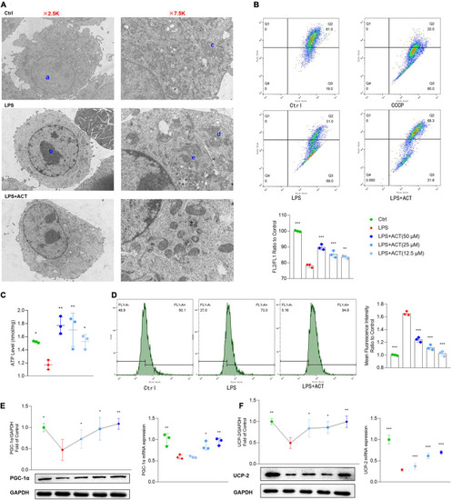
Effects of ACT on mitochondrial function in LPS-treated BV-2 cells. (A) Ultrastructural changes in the different groups of BV-2 cells under the transmission electron microscopy. a, normal nucleus. b, nucleus chromatin condensation. c, normal mitochondria. d, mitochondrial cristae disarranged or even disappeared. e, swollen and vacuolated mitochondria. (B) The representative images and bar graph of JC-1 red and green fluorescence ratio. (C) Bar graph showing quantified statistics of ATP level. (D) The representative images of DCFH-DA staining and bar graph of intracellular ROS production. (E) ACT distinctly decreases the protein and mRNA levels of PGC-1α. (F) ACT distinctly decreases the protein and mRNA levels of UCP-2. *P < 0.05, **P < 0.01, ***P < 0.005, versus the LPS group. n = 3.

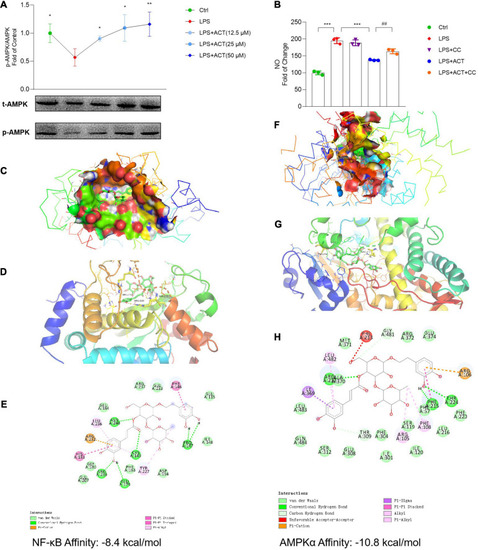
ACT bond to and inhibited NF-κB as well as activated AMPKα. (A) ACT distinctly increases the phosphorylation level of AMPKα. (B) The effect of compound C (1 μM) on NO level. ***P < 0.001, versus the LPS group. ##P < 0.01, versus the LPS+ACT group. n = 3. (C) A three-dimensional visual of the binding sites between ACT and NF-κB. (D) A two-dimensional close-up of the molecule binding pocket from the side. (E) Amino acid residues. (F) A three-dimensional visual of the binding sites between ACT and AMPKα. (G) A two-dimensional close-up of the molecule binding pocket from the side. (H) Amino acid residues.

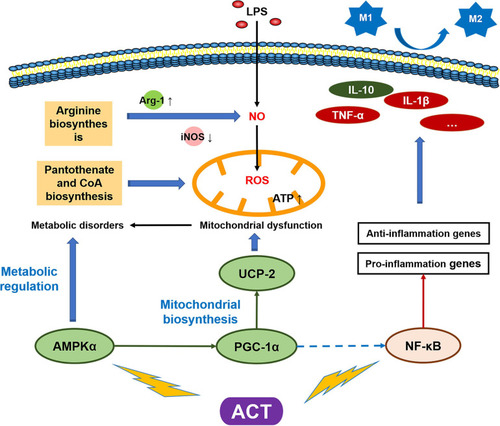
Schematic model of the mechanism by which ACT suppresses LPS-induced M1 polarization via regulating AMPK and NF-κB signaling pathways.

Acknowledgments
This image is the copyrighted work of the attributed author or publisher, and ZFIN has permission only to display this image to its users. Additional permissions should be obtained from the applicable author or publisher of the image. Full text @ Front Cell Dev Biol