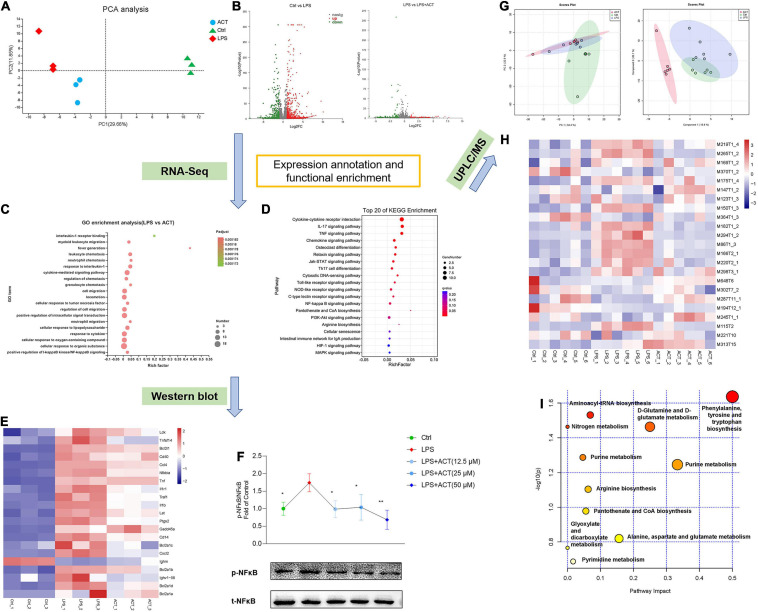
IMAGE

FIGURE 3

ID
ZDB-IMAGE-210807-5
Source
Figures for Li et al., 2021
Image
Figure Caption

FIGURE 3

ACT regulated M1/M2 polarization via the inhibition of NF-κB signaling pathway and positively regulated the metabolism shifts in LPS-induced BV-2 cells. (A) Principal components analysis (PCA) score plot for discriminating the cell metabolome from Ctrl, LPS, and ACT (50 μM) groups from a transcriptomic level (n = 3). (B) Volcano plot representing the RNA-seq data of LPS-induced and ACT-treated BV-2 cells. (C) GO terms in ACT-treated cells as determined by GO enrichment analysis. (D) Top 20 pathways in ACT-treated cells as determined by KEGG enrichment analysis. (E) Heat map of the differentially expressed genes (DEGs) that were linked to the NF-κB signaling pathway. (F) ACT distinctly decreased the phosphorylation level of NF-κB (n = 3). *P < 0.05, **P < 0.01, versus the LPS group. (G) PCA score plot and partial least squares-discrimination analysis (PLS-DA) score plot for discriminating the cell metabolome from Ctrl, LPS, and ACT (50 μM) groups in positive ion mode. (H) Heat map of the differential metabolites that were altered in the LPS group compared to the Ctrl group and the ACT group compared to the LPS group (n = 6). (I) The disturbed metabolic pathways in ACT group.

Acknowledgments
This image is the copyrighted work of the attributed author or publisher, and ZFIN has permission only to display this image to its users. Additional permissions should be obtained from the applicable author or publisher of the image. Full text @ Front Cell Dev Biol