
- Title
-
Immunohistochemical expression of aromatase cyp19a1a and cyp19a1b in the ovary and brain of zebrafish (Danio rerio) exposed to different concentrations of bisphenol A
- Authors
- Risalde, M.A., Molina, A.M., Lora, A.J., Ayala, N., Gómez-Villamandos, J.C., Moyano, M.R.
- Source
- Full text @ Aquat. Toxicol.
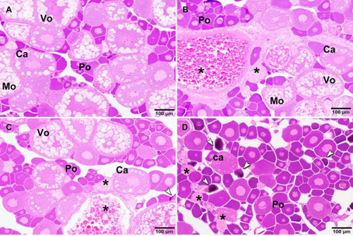
Histopathological changes in the zebrafish ovary exposed to different concentrations of BPA in water. A) Ovarian parenchyma of control zebrafish not exposed to BPA with numerous follicles at different stages of maturation, without histopathological lesions. B) Occasional presence of atretic follicles (asterisk) in ovary from zebrafish exposed to 10 μg/L of BPA. C) Presence of some atretic follicles (asterisk) and occasional presence of primordial follicles apoptosis (arrow) in ovary from zebrafish exposed to 100 μg/L of BPA. D) Multiple primary oocytes, primordial follicles apoptosis (arrows) and atretic follicles (asterisk) in ovary from zebrafish exposed to 1000 μg/L of BPA. Po-Primary oocyte; Ca-oocyte in cortical alveolus stage; Vo-vitellogenic oocyte; Mo-mature oocyte. Hematoxilin-eosine stain. PHENOTYPE:
|
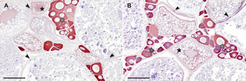
Cyp19a1a expression in the zebrafish ovary. Ooplasm of primary oocytes (gray asterisks) and peri-follicular cells of pre-vitellogenic and vitellogenic oocytes (arrows) were strongly stained against anti-cyp19a1a antibody, as well as lightly in ooplasm of vitellogenic oocyte of the ovary from zebrafish control (A) and exposed to 10 μg/L of BPA (B). Immunohistochemistry (ABC method). Bars: 100 μm. |
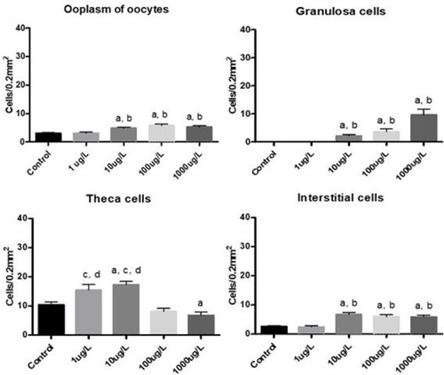
Response of cyp19a1a expression in the zebrafish ovary after exposure to different BPA concentrations. Means ± standard errors of the ooplasm of oocytes, as well as granulosa, theca and interstitial cells immunopositive for cyp19a1a in the ovary from control zebrafish and groups exposed to 1, 10, 100 and 1000 μg/L of BPA (n = 6 per group). asignificant differences vs control (p<0.05); bsignificant differences vs 1 μg/L (p<0.05); csignificant differences vs 100 μg/L (p<0.05); dsignificant differences vs 1000 μg/L (p<0.05) (Kruskal-Wallis test followed by Dunn's multiple comparisons test for non-parametric distributions). |
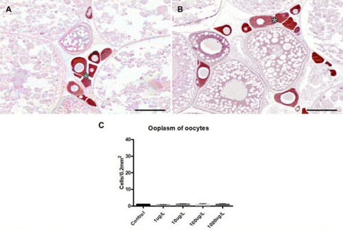
Cyp19a1b expression in the zebrafish ovary. Ooplasm of numerous primary oocytes was the main immunoreactive localization against anti-cyp19a1b antibody (asterisks) in the ovary of zebrafish control (A) and exposed to 10 μg/L of BPA (B). Immunohistochemistry (ABC method). Bars: 100 μm. C) Means ± standard errors of the ooplasm of oocytes immunopositive for cyp19a1b in the ovary from control zebrafish and groups exposed to 1, 10, 100 and 1000 μg/L of BPA (n = 6 per group). EXPRESSION / LABELING:
|
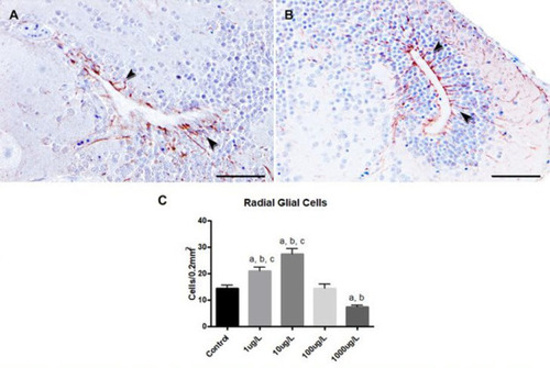
Response of cyp19a1b expression in the zebrafish brain after exposure to different BPA concentrations. The radial glial cells were the main immunoreactive cells against the anti-cyp19a1b antibody (arrows) in the brain from zebrafish control (A) and exposed to 10 μg/L of BPA (B). Immunohistochemistry (ABC method). Bars: 50 μm. C) Means ± standard errors of radial glial cells immunopositive for cyp19a1b in the brain from control zebrafish and groups exposed to 1, 10, 100 and 1000 μg/L of BPA (n = 6 per group). asignificant differences vs control (p<0.05); bsignificant differences vs 100 μg/L (p<0.05); csignificant differences vs 1000 μg/L (p<0.05) (Kruskal-Wallis test followed by Dunn's multiple comparisons test for non-parametric distributions). |