
Image
|
Figure Caption
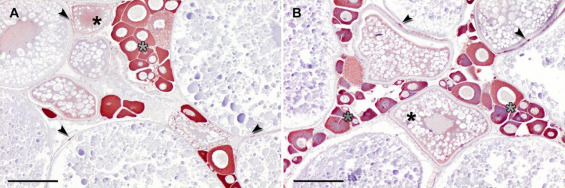
Fig. 2 Cyp19a1a expression in the zebrafish ovary. Ooplasm of primary oocytes (gray asterisks) and peri-follicular cells of pre-vitellogenic and vitellogenic oocytes (arrows) were strongly stained against anti-cyp19a1a antibody, as well as lightly in ooplasm of vitellogenic oocyte of the ovary from zebrafish control (A) and exposed to 10 μg/L of BPA (B). Immunohistochemistry (ABC method). Bars: 100 μm.
Figure Data
Acknowledgments
This image is the copyrighted work of the attributed author or publisher, and
ZFIN has permission only to display this image to its users.
Additional permissions should be obtained from the applicable author or publisher of the image.
Full text @ Aquat. Toxicol.