FIGURE

Fig. 5

ID
ZDB-FIG-220504-33
Publication
Risalde et al., 2021 - Immunohistochemical expression of aromatase cyp19a1a and cyp19a1b in the ovary and brain of zebrafish (Danio rerio) exposed to different concentrations of bisphenol A
Other Figures
All Figure Page
Back to All Figure Page
Fig. 5

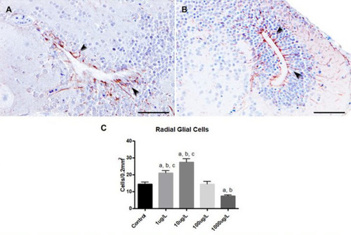
Response of cyp19a1b expression in the zebrafish brain after exposure to different BPA concentrations. The radial glial cells were the main immunoreactive cells against the anti-cyp19a1b antibody (arrows) in the brain from zebrafish control (A) and exposed to 10 μg/L of BPA (B). Immunohistochemistry (ABC method). Bars: 50 μm. C) Means ± standard errors of radial glial cells immunopositive for cyp19a1b in the brain from control zebrafish and groups exposed to 1, 10, 100 and 1000 μg/L of BPA (n = 6 per group). asignificant differences vs control (p<0.05); bsignificant differences vs 100 μg/L (p<0.05); csignificant differences vs 1000 μg/L (p<0.05) (Kruskal-Wallis test followed by Dunn's multiple comparisons test for non-parametric distributions).

Expression Data
Antibody:
Fish:
Condition:
Anatomical Term:
Stage: Adult

Expression Detail
Antibody Labeling
Phenotype Data
Fish:
Condition:
Observed In:
Stage: Adult

Phenotype Detail
Acknowledgments
This image is the copyrighted work of the attributed author or publisher, and ZFIN has permission only to display this image to its users. Additional permissions should be obtained from the applicable author or publisher of the image. Full text @ Aquat. Toxicol.