FIGURE SUMMARY
Title

Exploration of novel heterofused 1,2,4-triazine derivative in colorectal cancer

Authors
Hermanowicz, J.M., Szymanowska, A., Sieklucka, B., Czarnomysy, R., Pawlak, K., Bielawska, A., Bielawski, K., Kalafut, J., Przybyszewska, A., Surazynski, A., Rivero-Muller, A., Mojzych, M., Pawlak, D.
Source
Full text @ J Enzyme Inhib Med Chem

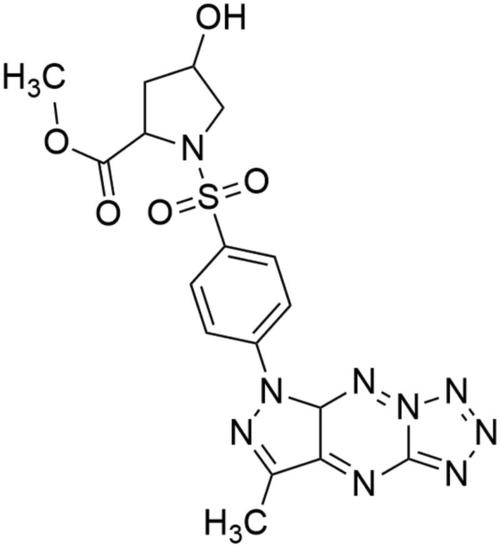
Chemical structure of MM-129.

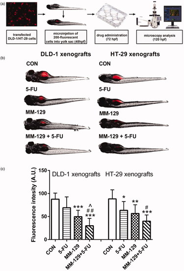
Schematic of xenograft assay and analysis of tumour development (a). Site-specific injection (yolk sac) of transfected (red) colon cancer cells (DLD-1 and HT-29) into 48 hpf zebrafish embryos and imaging analysis of tumour growth after 48 h of incubation with MM-129 (50 µM), 5-FU (50 µM), or a combination of these agents (b). Quantification of total fluorescence by colon cancer cells three days after injection (c) n = 8, *p < .05, **p < .01, ***<.001 vs. CON; ^p < .05 vs. MM-129, #p < .05, ##p < .01 vs. 5-FU. Data were presented as mean ± standard deviation (SD), and analysed using one-way analysis of variance (ANOVA). p < .05 was considered statistically significant.

Effect of MM-129, 5-fluorouracil (5-FU), and roscovitine (RSC) on cell division in the zebrafish embryo. Zebrafish eggs after 0, 1, 1.25, and 2 h of exposure to MM-129, 5-FU and RSC; n = 10. hpf: hours post fertilisation; hpt: hours post treatment.

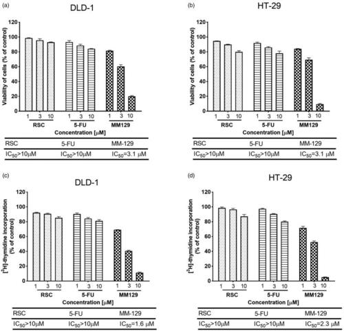
Viability of DLD-1 (a) and HT-29 (b) colon cancer cells treated for 24 h with different concentrations of MM-129, roscovitine (RSC) and 5-fluorouracil (5-FU), and the antiproliferative effects of the studied compounds in DLD-1 (c) and HT-29 (d) colon cancer cells measured by the inhibition of [3H]thymidine incorporation into DNA. Mean values ± SD from three independent experiments (n = 3) done in duplicate are presented.

The down-regulation of Bruton’s kinase (BTK) expression by MM-129. Phosphorylated BTK (pBTK) expression was determined by Western blot (a) and confocal microscopy (d) in DLD-1 and HT-29 cells treated with MM-129, roscovitine and 5-flurouracil in two concentrations: 1 µM and 3 µM for 24 h. The band staining was quantified by densitometry (b, c). Samples used for electrophoresis consisted of 20 µg of protein from six pooled cell extracts (n = 6). Data were presented as mean ± standard deviation (SD), and analysed using one-way analysis of variance (ANOVA). ***p<.001 vs. CON intensity of cytoplasmic/nuclear fluorescence was analysed in cell populations. Images of cells labelled with FITC were obtained using a 515LP emission laser and a 488/10 excitation laser (d).

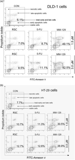
Representative flow cytometry dot-plots for annexin V‐FITC-propidium iodide assay of DLD-1 (a) and HT-29 (b) colon cancer cells after 24 h of incubation with roscovitine (RSC), 5-fluorouracil (5-FU), or MM-129 (1 μM and 3 μM).

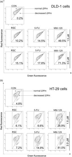
Representative dot-plots presenting the loss of mitochondrial membrane potential (ΔΨm) of DLD-1 (a) and HT-29 (b) colon cancer cells treated for 24 h with roscovitine (RSC), 5-fluorouracil (5-FU), and MM-129 (1 μM and 3 μM).

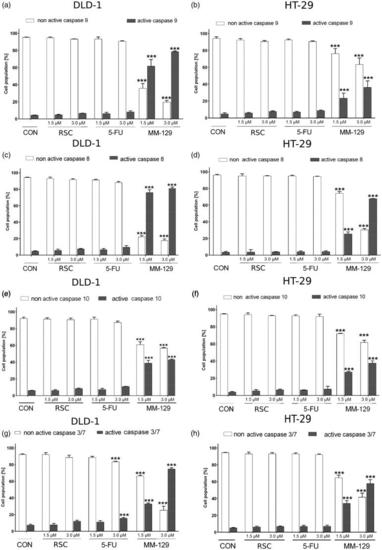
Flow cytometric analysis of caspase-9, caspase-8, caspase-10, and caspases-3/7 activation in the populations of DLD-1 (a, c, e, g) and HT-29 (b, d, f, h) colon cancer cells treated for 24 h with roscovitine (RSC), 5-fluorouracil (5-FU), and MM-129 (1 μM and 3 μM). Mean percentage values from three independent experiments done in duplicate (N = 6) were presented as mean ± standard deviation (SD), and analysed using one-way analysis of variance (ANOVA). ***p<.001 vs. CON.

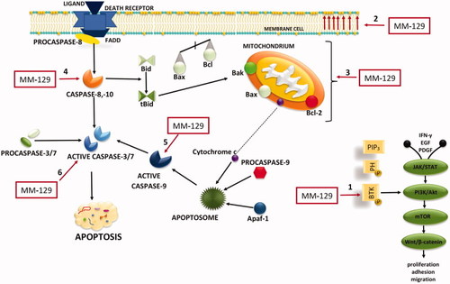
Schematic representation of possible anticancer mechanisms of MM-129. 1: BTK inhibition; 2: phosphatidylserine (PS) externalisation; 3: loss of mitochondrial membrane potential; 4: activation of extrinsic pathway of apoptosis; 5: the activation of internal (mitochondrial) apoptosis; 6: activation of executive caspases. The schematic illustration was created in Adobe Photoshop and Photophea software. Akt: protein kinase B; Apaf-1: apoptotic protease activating factor 1; β-catenin: protein responsible for transduction; Bak: Bcl-2 homologous antagonist/killer; Bax: Bcl-2-associated X protein; Bcl-2: antiapoptotic protein; Bid: Bax-like BH3 protein; tBid: truncated BID; BTK: Bruton’s tyrosine kinase; EGF: epidermal growth factor; FADD: Fas-associated death domain protein; IFNγ: interferon gamma; JAK2: non-receptor tyrosine kinase; mTOR: mammalian target of rapamycin; PDGF: platelet-derived growth factor; PI3K: phosphoinositide 3-kinases; PIP3: phosphatidylinositol-3,4,5-triphosphate; PH: pleckstrin homology domain; STAT: signal transducer and activator of transcription; Wnt: family of secreted lipid-modified signalling glycoproteins.

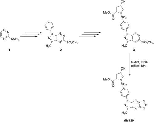
Synthetic pathway for production of new sulphonamide MM-129.

Acknowledgments
This image is the copyrighted work of the attributed author or publisher, and ZFIN has permission only to display this image to its users. Additional permissions should be obtained from the applicable author or publisher of the image. Full text @ J Enzyme Inhib Med Chem