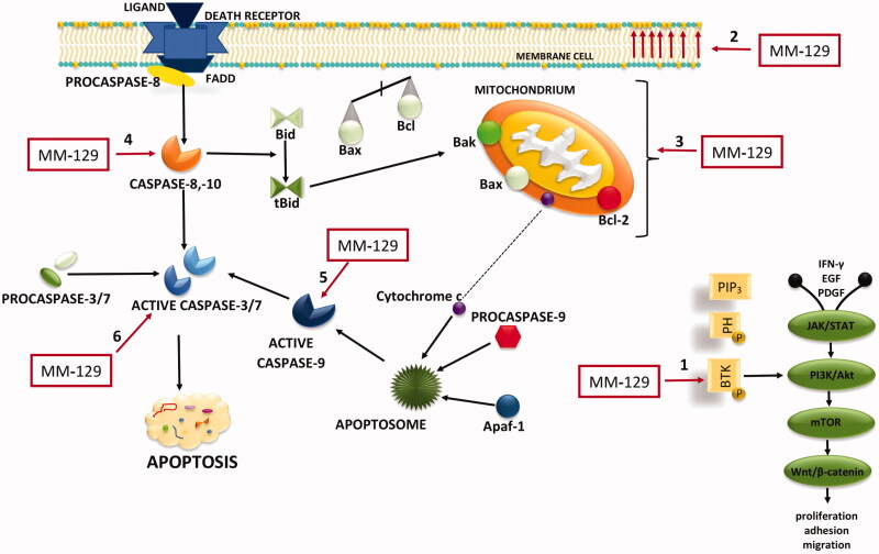
IMAGE

Figure 9.

ID
ZDB-IMAGE-210208-40
Source
Figures for Hermanowicz et al., 2021
Image
Figure Caption

Figure 9.

Schematic representation of possible anticancer mechanisms of MM-129. 1: BTK inhibition; 2: phosphatidylserine (PS) externalisation; 3: loss of mitochondrial membrane potential; 4: activation of extrinsic pathway of apoptosis; 5: the activation of internal (mitochondrial) apoptosis; 6: activation of executive caspases. The schematic illustration was created in Adobe Photoshop and Photophea software. Akt: protein kinase B; Apaf-1: apoptotic protease activating factor 1; β-catenin: protein responsible for transduction; Bak: Bcl-2 homologous antagonist/killer; Bax: Bcl-2-associated X protein; Bcl-2: antiapoptotic protein; Bid: Bax-like BH3 protein; tBid: truncated BID; BTK: Bruton’s tyrosine kinase; EGF: epidermal growth factor; FADD: Fas-associated death domain protein; IFNγ: interferon gamma; JAK2: non-receptor tyrosine kinase; mTOR: mammalian target of rapamycin; PDGF: platelet-derived growth factor; PI3K: phosphoinositide 3-kinases; PIP3: phosphatidylinositol-3,4,5-triphosphate; PH: pleckstrin homology domain; STAT: signal transducer and activator of transcription; Wnt: family of secreted lipid-modified signalling glycoproteins.

Acknowledgments
This image is the copyrighted work of the attributed author or publisher, and ZFIN has permission only to display this image to its users. Additional permissions should be obtained from the applicable author or publisher of the image. Full text @ J Enzyme Inhib Med Chem