FIGURE SUMMARY
Title

Tetraspanin18 regulates angiogenesis through VEGFR2 and Notch pathways

Authors
Li, G.X., Zhang, S., Liu, R., Singh, B., Singh, S., Quinn, D.I., Crump, G., Gill, P.S.
Source
Full text @ Biol. Open

Tspan18 expression in the cardiovascular system. The expression of Tspan18 and its paralogue Tspan12 was analyzed by RT-PCR in (A) human fetal tissues and (B) primary cultured endothelial cells and tumor cell lines. UASMC, umbilical artery smooth muscle cell; HUVEC, human umbilical vein endothelial cell; HUAEC, human umbilical artery endothelial cell; NC, negative control with no cDNA. (C) Quantitative RT-PCR analysis of Tspan18 expression at different times in zebrafish development (whole embryo). Expression was normalized to β–actin. (D) Whole-mount ISH of Tspan18 expression in the developing embryos. D is the cross section of C. DA, dorsal aorta; PCV, posterior cardinal vein; DLAV, dorsal longitudinal anastomotic vessels; ISV, intersegmental vessel; S, somites; NC, Notochord. All larvae lateral view unless indicated. Anterior is to the left. Scale bars:100 μm. All experiments (A–D) were repeated at least three times and similar results were obtained.

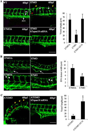
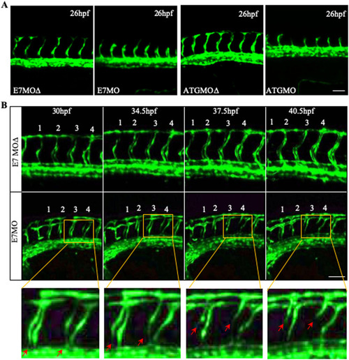
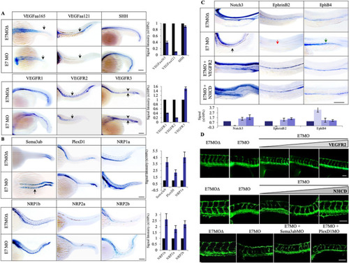
Vascular defects in Tspan18-zebrafish morphants. (A) Morphological and vascular defects were examined in E7MO-injected zebrafish at 60 hpf. E7MO represents the morpholino targeting Tspan18 Exon 7 splicing site. E7MOΔ is a control morpholino with five-base mismatch. WT represents fish injected with morpholino diluent only. Arrows in the picture of Tspan18-deficient fish (E7MO) point to missing ISV (red), ectopic ISV branching (white) or discontinuous DLAV (yellow). Quantification of ISV length of E7MO and E7MOΔ injected fish (n=80) are shown on the bottom left. Co-injection of a human sense capped Tspan18 mRNA rescued the defects caused by E7MO. (B) E7MOΔ injected embryos had reduced subintestinal vessels (upper panel, arrows) and narrow ISV lumen (lower panel, arrowheads) at 72 hpf. Quantification of ISV lumen width from each group is shown on the right (n=75). (C) Morphological and vascular defects were examined in ATGMO injected zebrafish at 60 hpf. ATGMO represents the morpholino targeting Tspan18 ATG start codon containing Exon 3 splicing site. Arrows are designated as in A. Co-injection of a human sense capped Tspan18 mRNA rescued the defects caused by ATGMO. Error bars represent s.d. P value was calculated using two-tailed Student's t-test. *P<0.02. All larvae lateral view. Anterior is to the left. Scale bars: 50 μm. All experiments (A–C) were repeated at least three times and similar results were obtained.

Vessel regression in Tspan18 zebrafish morphants. (A) Intact ISV sprouting is observed at 26 hpf in Tspan18-deficient fish. (B) Confocal time-lapse microscopy of regressing ISVs (red arrows) in Tspan18-deficient fish. Numbers 1–4 indicate the order of ISVs. Yellow rectangle: corresponding section shown at higher magnification under the bottom. All larvae lateral view. Anterior is to the left. Scale bars: 50 μm. All experiments (A–B) were repeated at least three times and similar results were obtained.

A cartoon illustrating pathways involved in zebrafish embryonic angiogenesis. SHH is a master regulator of angiogenesis and it regulates VEGF/VEGFR2. Neuropilin as a co-receptor is important for VEGF-VEGFR2 interaction and signaling, and regulates Semaphorin-PlexinD1 signaling. VEGFR2 signaling leads to vessel sprouting, whereas PlexinD1 signaling restricts angiogenesis and regulates vessel patterning. VEGFR2 induces regulates Dll4/Notch signaling, which in turn determines vessel specification (EphB4 on vein and EphrinB2 on artery) and vessel stability through Nrarp.

Tspan18 functions through VEGF/VEGFR and Notch signaling pathways. (A) Whole-mount ISH analysis of SHH, VEGFs and VEGFRs at 27 hpf in E7MO injected zebrafish. The expression of VEGFs especially VEGFaa121 and VEGFR2 was decreased (indicated by arrows) while the expression of VEGFR3 was increased in PCV (arrows) and ectopically expressed in DA (arrowheads). No significant changes were observed in SHH and VEGFR1 expression. Quantification of each genes is shown on the right (n>10). (B) Whole-mount ISH analysis of Sema3ab, PlexD1 and four Nrp isoforms in E7MO injected fish at 27 hpf. Sema3ab is highly induced in anterior somites (arrow). Quantification of each genes is shown on the right (n>10). (C) Whole-mount ISH analysis of Notch3, EphrinB2, and EphB4 at 27 hpf. In Tspan18 deficient fish, Notch3 (black arrow) and EphrinB2 (red arrow) were lost in the DA, while EphB4 was ectopically expressed in the DA (green arrow). These defects were rescued by co-injection of zebrafish VEGFR2 sense capped mRNA or Notch3 intracellular domain (N3ICD) mRNA. Quantification of each gene is shown at the bottom (n>10). (D) VEGFR2 (top) and N3ICD (middle) mRNA rescued E7MO-morphants in a dose-dependent manner (1.0, 1.5 and 2.0 ng per injection), whereas MOs targeting Sema3ab and PlexD1 (bottom) only partially rescued the defects (6 and 10 ng per injection respectively). Images of 27 hpf zebrafish are shown. Similar results were obtained from three independent experiments. All larvae lateral view. Anterior is to the left. Scale bars: 100 μm.

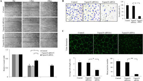
Tspan18 knockdown inhibits angiogenesis in vitro. (A) Tspan18 knockdown inhibited migration of HUAEC in an in vitro wound-healing assay. Transfection reagents served as a negative control. The margin of the wound is demarcated transwelled lines. Three independent experiments were performed and quantified wound width is shown (normalized to 0 h width). (B) Tspan18 knockdown inhibited invasion of HUAEC in transwell invasion assay. Quantification of invaded cells is shown on the right. (C) Tspan18 knockdown inhibited HUAEC tube formation on Matrigel. Quantification of tube length and number of junctions is shown on the bottom. The experiments in A–C were repeated three times with similar results. Error bars: s.d., *P<0.01, **P<0.002. P values were calculated using a two-tailed Student's t-test.

Knockdown of Tspan18 by siRNA leads to downregulation of VEGFR2 and Notch1 in vitro. (A) Western blot analysis of VEGFR2 and VEGFR1 expression in Tspan18 siRNA transfected HUAEC. A mutant siRNA (Tspan18 siRNAΔ) was used as siRNA control. β-actin served as loading control. (B) VEGFR2 and hNICD normalized the gene-expression changes in Tspan18-deficient HUAEC. VEGFR2 and hNICD were overexpressed in HUAEC transfected with Tspan18 siRNA. Gene expression was analyzed by quantitative RT-PCR, normalized to β-actin level and compared to that of Tspan18 siRNAΔ-treated HUAEC (set as 1.0). The experiments were repeated three times with similar results. Error bars: s.d., *P<0.01, **P<0.002. P values were calculated using a two-tailed Student's t-test. (C) Western blots showing the downregulation of Notch1 and VEGFR2 in VEGFR2-expressing mesothelioma cell line 211H by Tspan18 siRNA. No change in Notch1 is observed in cervical cancer Hela cell line that has no VEGFR2. This experiment was repeated three times with similar results. The immunoblots were quantified with Image J and the relative band intensity is shown.

Acknowledgments
This image is the copyrighted work of the attributed author or publisher, and ZFIN has permission only to display this image to its users. Additional permissions should be obtained from the applicable author or publisher of the image. Full text @ Biol. Open