FIGURE SUMMARY
Title

Ethyl Acetate Fraction of Aqueous Extract of Lentinula edodes Inhibits Osteoclastogenesis by Suppressing NFATc1 Expression

Authors
Lee, H., Lee, K., Lee, S., Lee, J., Jeong, W.T., Lim, H.B., Hyun, T.K., Yi, S.J., Kim, K.
Source
Full text @ Int. J. Mol. Sci.

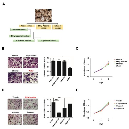
Ethyl acetate fraction of aqueous extract of Lentinula edodes inhibits RANKL-mediated osteoclastogenesis. (A) Schematic representation showing the extraction and fractionation of anti-osteoclastic compounds from L. edodes. (B) BMMs were treated with M-CSF (30 ng/mL) and RANKL (100 ng/mL) in the presence or absence of 10 μg/mL three extract (water extract, ethyl acetate extract, ethanol extract) of L. edodes for three days, and osteoclast formation was examined by tartrate-resistant acid phosphatase (TRAP) staining (left) and counting the number of TRAP-positive multinuclear osteoclasts (right). (C) BMMs were treated with M-CSF (30 ng/mL) and RANKL (100 ng/mL) in the presence of the extracts as in (B), and cell proliferation was measured by MTT assays. (D) BMMs were treated with M-CSF (30 ng/mL) and RANKL (100 ng/mL) in the presence of four fractions (10 μg/mL) of water extract of L. edodes for three days, and osteoclast differentiation was assessed by TRAP staining (left) and the number of TRAP-positive multinuclear osteoclasts (right) was counted. (E) BMMs were treated with the fractions as in (D), and cell proliferation was measured by MTT assays. Error bars represent the mean result ± SD of three independent experiments; * p < 0.05, *** p < 0.001.

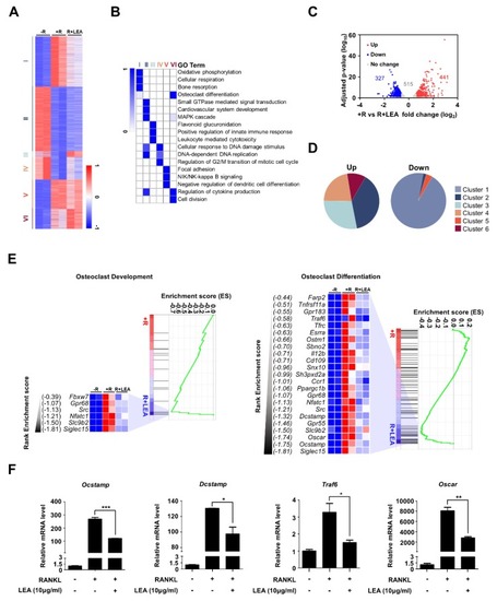
LEA alters gene expression profiling in BMMs. (A) K-means (K = 6) clustering of 4740 differentially expressed genes (DEGs) in any pairwise comparison among three conditions (−R; no RANKL, +R; RANKL, R + LEA; ethyl acetate fraction of L. edodes with RANKL). Clusters are indicated on the left. (B) Heatmap showing the p-value significance of GO term enrichment for genes in each cluster. (C) Volcano plot of transcriptomic changes between +R and R + LEA. Genes with increased (red) or decreased (blue) expression in LEA + RANKL-treated cells relative to RANKL-treated cells were defined based on FDR-adjusted p < 0.05 and greater than 1.5-fold expression changes. (D) Pie charts showing each cluster portion of as in (C). Up: Cluster 1 (0%), Cluster 2 (38.5%), Cluster 3 (28.3%), Cluster 4 (22.2%), Cluster 5(0%), and Cluster 6 (11%). Down: Cluster 1 (94.5%), Cluster 2 (1.8%), Cluster 3 (0%), Cluster 4 (0%), Cluster 5 (3.7%), and Cluster 6 (0%). (E) GSEA of 4740 genes as in (A) shows the enrichment of genes associated with osteoclast development and osteoclast differentiation. Heatmap represents core enriched genes based on their enrichment score. (F) qRT-PCR was performed to quantify relative mRNA levels of the representative genes for osteoclastogenesis. Error bars represent the mean result ± SD of three independent experiments; * p < 0.05, ** p < 0.01, *** p < 0.001.

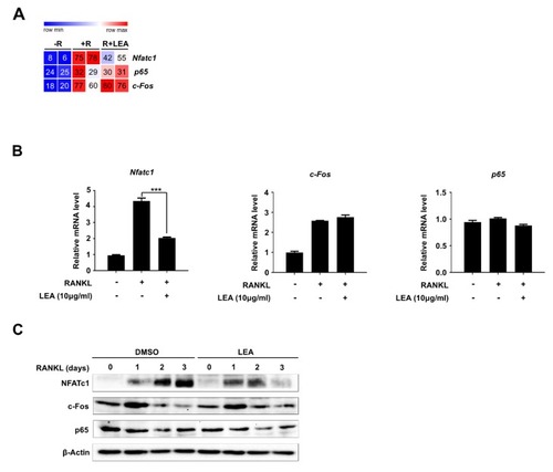
LEA represses RANKL-mediated NFATc1 expression. (A) Heatmap showing the reads per kilobase per million (RPKM) of c-Fos, NF-κB and NFATc1. Numbers within the figure indicate RPKM values. (B) Total RNA was prepared after treated with M-CSF (30 ng/mL) and RANKL (100 ng/mL) in the presence or absence of LEA (10 μg/mL), and qRT-PCR was performed using primers specific for Nfatc1, c-Fos, and p65. Results represents the means ± SD of three independent experiments. *** p < 0.001 versus only RANKL treatment. (C) Whole cell lysates were prepared from M-CSF/RANKL-treated BMMs with or without LEA (10 μg/mL) for 0, 1, 2, and 3 days, and analyzed by immunoblotting with NFATc1, c-Fos and p65 antibodies. β-Actin was probed as a loading control.

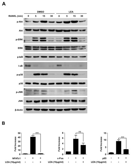
LEA suppresses NFATc1 expression by blocking both NF-κB and NFATc1-mediated transactivity. (A) BMMs were pre-treated with DMSO and LEA (10 μg/mL) for 1 h, and stimulated with RANKL for 0, 5, 15, and 30 min. The cells were lysed and immunoblotted with antibodies against p-Akt, Akt, p-ERK, ERK, p-IκB, IκB, p-p38, p38, p-JNK, and JNK. Band intensities were normalized to β-Actin control. (B) 293T cells were transiently transfected with the reporter plasmid Nfatc1-Luc along with c-Fos, p65, or Nfatc1 in the presence or absence of LEA (10 μg/mL). Luciferase activity was measured after 24 h post-transfection using Luciferase assay kit (Promega, Madison, WI, USA). Each bar represents the means ± S.D. of three independent experiments. *** p < 0.001. ns = not significant.

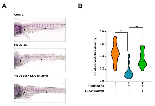
Anti-osteoporotic effect of LEA in a model of prednisolone-induced osteoporosis in zebrafish. (A) The larvae at 10 dpf (days post-fertilization) were treated with 25 µM prednisolone (PS) in the presence or absence of LEA (10 μg/mL) for 3 days. Whole-mount Alizarin red staining was performed to analyze the mineralized bone. Arrowheads indicate the bone Alizarin red staining. (B) Relative vertebral bone density was assessed by measuring the areas of the first ten stained vertebrae (V1–V10, indicated by arrowhead in (A)). *** p < 0.001 versus only prednisolone treatment.

Acknowledgments
This image is the copyrighted work of the attributed author or publisher, and ZFIN has permission only to display this image to its users. Additional permissions should be obtained from the applicable author or publisher of the image. Full text @ Int. J. Mol. Sci.