
- Title
-
Axonal regeneration in zebrafish spinal cord
- Authors
- Ghosh, S., Hui, S.P.
- Source
- Full text @ Regeneration (Oxf)
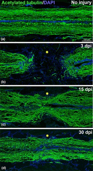
Time course of axonal regeneration in adult zebrafish stained with acetylated‐tubulin (green) and DAPI (blue). (A) Uninjured adult spinal cord. (B) 3‐day post transected spinal cord showing complete loss of axonal connections (green) in the injury epicenter (yellow star). (C) 15‐day post transected spinal cord showing some regenerated axons passing through the injury epicenter (yellow star). (D) A 30‐day post transected spinal cord showing significant numbers of regenerated axons passing through the injury epicenter (yellow star). Significant axonal regrowth can be observed compared to uninjured cord. All the images are of the same magnification. Scale bar 100 μm (A, B, C, and D) |
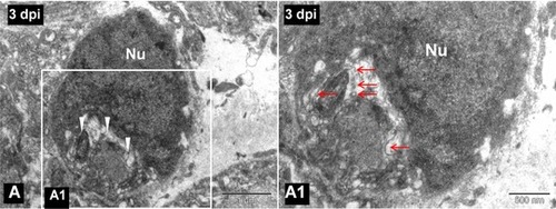
(A) TEM image of a 3‐day post injured spinal cord showing a phagocytic macrophage as cytoplasm is filled with myelin debris (white arrowheads). (A1) Higher magnification image of the boxed area in (A), in which degenerated myelin debris (red arrows) is clearly visible in the cytoplasm of the same phagocytic macrophage. Nu, cell nucleus. Scale bar 1 μm (A), 500 nm (A1) |
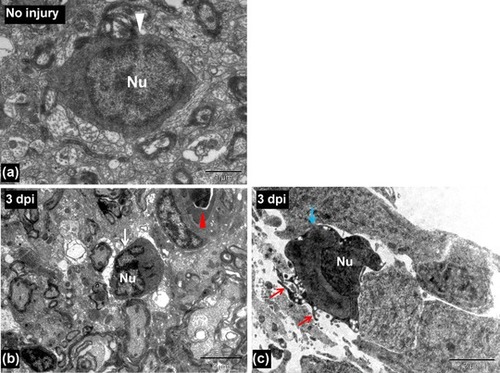
(A) TEM image of an uninjured spinal cord showing a microglia (white arrowhead). (B) TEM image of a 3‐day post injured spinal cord showing a microglia (white arrow) near the injury site. Red arrowhead indicates a blood vessel. (C) TEM image of a 3‐day post injured spinal cord showing an activated microglia near the central canal (having finger‐like cytoplasmic projections, red arrows) of injured spinal cord (blue arrow). Nu, cell nucleus. Scale bar 1 μm (A), 5 μm (B), 2 μm (C) |
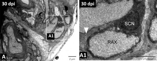
(A) TEM image of 30‐day regenerated spinal cord showing remyelinating axons wrapped by a Schwann cell. (A1) Higher magnification image of the boxed area in (A), in which the white arrowhead indicates the basal membrane of the Schwann cell, remyelinating an axon. SCN, nucleus of the Schwann cell; RAX, regenerating axon. Scale bar 2 μm (A), (A1). Adapted from Hui et al., |