
Figure 1
- ID
- ZDB-FIG-190723-2444
- Publication
- Ghosh et al., 2018 - Axonal regeneration in zebrafish spinal cord
- Other Figures
- All Figure Page
- Back to All Figure Page
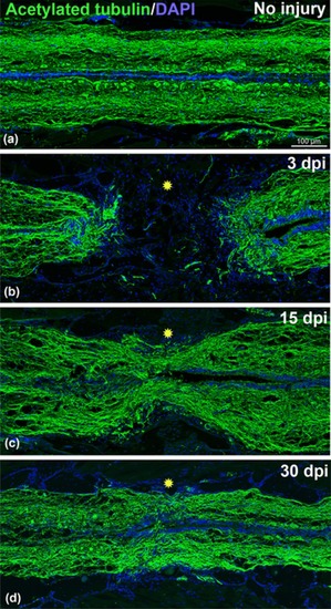
Time course of axonal regeneration in adult zebrafish stained with acetylated‐tubulin (green) and DAPI (blue). (A) Uninjured adult spinal cord. (B) 3‐day post transected spinal cord showing complete loss of axonal connections (green) in the injury epicenter (yellow star). (C) 15‐day post transected spinal cord showing some regenerated axons passing through the injury epicenter (yellow star). (D) A 30‐day post transected spinal cord showing significant numbers of regenerated axons passing through the injury epicenter (yellow star). Significant axonal regrowth can be observed compared to uninjured cord. All the images are of the same magnification. Scale bar 100 μm (A, B, C, and D) |