
FIGURE
Figure 2
- ID
- ZDB-FIG-190723-2445
- Publication
- Ghosh et al., 2018 - Axonal regeneration in zebrafish spinal cord
- Other Figures
- All Figure Page
- Back to All Figure Page
Figure 2
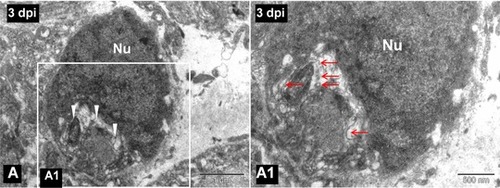
(A) TEM image of a 3‐day post injured spinal cord showing a phagocytic macrophage as cytoplasm is filled with myelin debris (white arrowheads). (A1) Higher magnification image of the boxed area in (A), in which degenerated myelin debris (red arrows) is clearly visible in the cytoplasm of the same phagocytic macrophage. Nu, cell nucleus. Scale bar 1 μm (A), 500 nm (A1) |
Expression Data
Expression Detail
Antibody Labeling
Phenotype Data
Phenotype Detail
Acknowledgments
This image is the copyrighted work of the attributed author or publisher, and
ZFIN has permission only to display this image to its users.
Additional permissions should be obtained from the applicable author or publisher of the image.
Full text @ Regeneration (Oxf)