
Image
|
Figure Caption
Figure 4
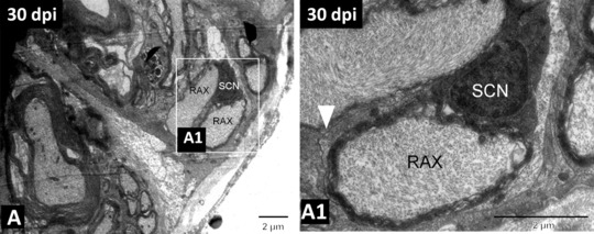
(A) TEM image of 30‐day regenerated spinal cord showing remyelinating axons wrapped by a Schwann cell. (A1) Higher magnification image of the boxed area in (A), in which the white arrowhead indicates the basal membrane of the Schwann cell, remyelinating an axon. SCN, nucleus of the Schwann cell; RAX, regenerating axon. Scale bar 2 μm (A), (A1). Adapted from Hui et al.,
Acknowledgments
This image is the copyrighted work of the attributed author or publisher, and
ZFIN has permission only to display this image to its users.
Additional permissions should be obtained from the applicable author or publisher of the image.
Full text @ Regeneration (Oxf)